. 2008 Mar;118(3):1056-64.
doi: 10.1172/JCI33871.

Increased expression of the WNT antagonist sFRP-1 in glaucoma elevates intraocular pressure

Affiliations

Increased expression of the WNT antagonist sFRP-1 in glaucoma elevates intraocular pressure

Wan-Heng Wang et al. J Clin Invest. 2008 Mar.

Abstract

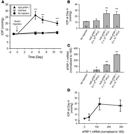
Elevated intraocular pressure (IOP) is the principal risk factor for glaucoma and results from excessive impedance of the fluid outflow from the eye. This abnormality likely originates from outflow pathway tissues such as the trabecular meshwork (TM), but the associated molecular etiology is poorly understood. We discovered what we believe to be a novel role for secreted frizzled-related protein-1 (sFRP-1), an antagonist of Wnt signaling, in regulating IOP. sFRP1 was overexpressed in human glaucomatous TM cells. Genes involved in the Wnt signaling pathway were expressed in cultured TM cells and human TM tissues. Addition of recombinant sFRP-1 to ex vivo perfusion-cultured human eyes decreased outflow facility, concomitant with reduced levels of beta-catenin, the Wnt signaling mediator, in the TM. Intravitreal injection of an adenoviral vector encoding sFRP1 in mice produced a titer-dependent increase in IOP. Five days after vector injection, IOP increased 2 fold, which was significantly reduced by topical ocular administration of an inhibitor of a downstream suppressor of Wnt signaling. Thus, these data indicate that increased expression of sFRP1 in the TM appears to be responsible for elevated IOP in glaucoma and restoring Wnt signaling in the TM may be a novel disease intervention strategy for treating glaucoma.


Figures

Figure 1
Figure 1. Identification of sFRP1 differential expression in glaucomatous TM cells.
RDD of normal TM cells compared with glaucomatous TM cells. M, molecular size markers; N1–N3, normal TM cells (NTM70A); G, glaucoma TM cells (GTM29A); L, control sample derived from human liver. Bold arrows indicate a differential display band in the glaucoma TM cells. Experiments 1 and 2 represent independent PCR reactions, which used the same samples and primers (HAP1/T11A). The DNA sequence of the differentially expressed cDNA fragment in glaucomatous TM cells is identical to sFRP1 (GenBank accession number NM_003012).
Figure 2
Figure 2. Confirmation of sFRP-1 differential expression in glaucomatous TM cells.
(A) Quantitative RT-PCR quantitation of sFRP-1 expression in 6 age-matched pairs of normal and glaucomatous TM cell lines. (B) ELISA analysis of sFRP-1 protein levels in 13 normal and 10 glaucomatous TM cell lines. Data are shown as mean ± SEM. *P < 0.05; **P < 0.01.
Figure 3
Figure 3. Effects of sFRP-1 on perfusion-cultured human eyes.
(A) Effect of recombinant human sFRP-1 (10 μg/ml) on aqueous outflow rate. The treatment started from time 0. *P < 0.05 versus same time point of the vehicle group (mean ± SEM; n = 4). There was no significant difference in basal flow rates between the 2 groups at time 0. (B) Western blot analysis of sFRP-1 and β-catenin in TM and CB of perfused human eyes. (C) ELISA assessment of β-catenin levels in control and sFRP-1–perfused TM and CB (mean ± SEM, n = 4). **P < 0.01.
Figure 4
Figure 4. Effects of Ad5.
FRP-1 on mouse IOP and sFRP1 expression. (A) Ad5.sFRP1 (3 × 10⁷ PFU/eye, n = 6) or Ad5.Null (3 × 10⁷ PFU/eye, n = 6) was injected intravitreally at day 0. IOP was measured with the TonoLab rebound tonometer. (B) Mouse IOP at day 4 after intravitreal injection of Ad5.sFRP-1 (2 × 10⁶, 1 × 10⁷, or 5 × 10⁷ PFU/eye). Sample sizes are indicated by the number at the base of each bar. (C) Expression of sFRP1 assayed by quantitative RT-PCR in mouse eyes injected with the vectors indicated in B at day 6 after injection. (D) Correlation between sFRP1 expression level and IOP in mice. Data are shown as mean ± SEM. *P < 0.05, **P < 0.01, ***P < 0.001 versus “No injection” group by 1-way ANOVA then Bonferroni’s test.
Figure 5
Figure 5. Effect of GSK inhibitor on sFRP1-induced ocular hypertension in mice.
After intraocular injection of Ad5.sFRP-1 (3 × 10⁷ PFU/eye) at day 0, twice daily topical ocular administrations of a GSK inhibitor, Compound 12 (1% wt/vol suspension), or vehicle were performed on days 3, 4, and 5 (gray horizontal bar). *P < 0.05 versus the “No injection” group; #P < 0.05 versus the “Ad5.sFRP-1 + Vehicle” group by 1-way ANOVA then Bonferroni’s test (mean ± SEM; n = 10).

Similar articles

Cited by

References

    1. Quigley H.A. Number of people with glaucoma worldwide. Br. J. Ophthalmol. 1996;80:389–393. - PMC - PubMed
    1. Quigley H.A. The search for glaucoma genes — implications for pathogenesis and disease detection. N. Engl. J. Med. 1998;338:1063–1064. - PubMed
    1. Quigley H.A. Open-angle glaucoma. N. Engl. J. Med. 1993;328:1097–1106. - PubMed
    1. Kass M.A., et al. 2002The Ocular Hypertension Treatment Study: a randomized trial determines that topical ocular hypotensive medication delays or prevents the onset of primary open-angle glaucoma. Arch. Ophthalmol. 120701–713; discussion 829–830. . - PubMed
    1. The AGIS Investigators. . The Advanced Glaucoma Intervention Study (AGIS): 7. The relationship between control of intraocular pressure and visual field deterioration.The AGIS Investigators. Am. J. Ophthalmol. 2000;130:429–440. - PubMed

Publication types

MeSH terms