. 2008 Apr;129(4):421-40.
doi: 10.1007/s00418-008-0396-9. Epub 2008 Feb 15.

The peroxisome: still a mysterious organelle

Affiliations

The peroxisome: still a mysterious organelle

Michael Schrader et al. Histochem Cell Biol. 2008 Apr.

Abstract

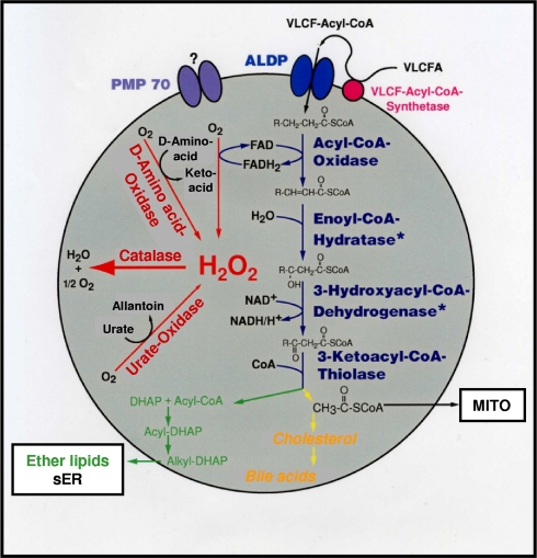
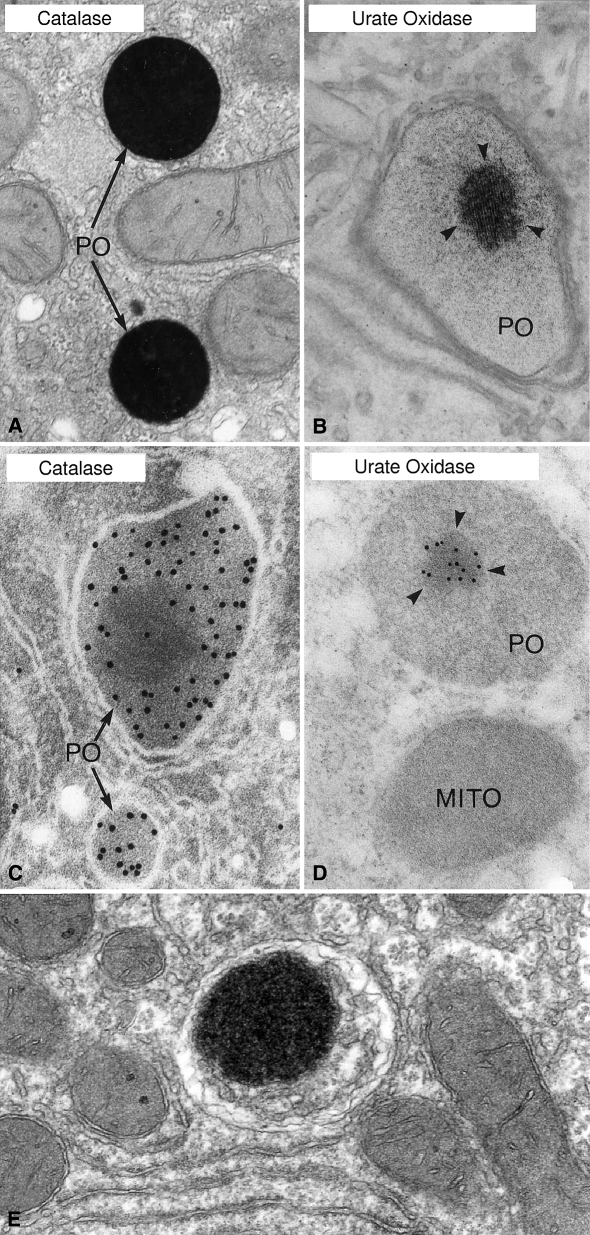
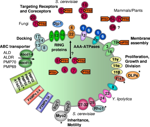
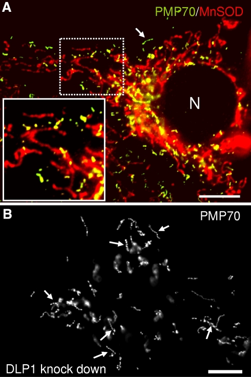
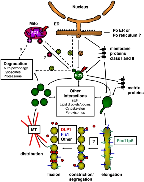
More than half a century of research on peroxisomes has revealed unique features of this ubiquitous subcellular organelle, which have often been in disagreement with existing dogmas in cell biology. About 50 peroxisomal enzymes have so far been identified, which contribute to several crucial metabolic processes such as beta-oxidation of fatty acids, biosynthesis of ether phospholipids and metabolism of reactive oxygen species, and render peroxisomes indispensable for human health and development. It became obvious that peroxisomes are highly dynamic organelles that rapidly assemble, multiply and degrade in response to metabolic needs. However, many aspects of peroxisome biology are still mysterious. This review addresses recent exciting discoveries on the biogenesis, formation and degradation of peroxisomes, on peroxisomal dynamics and division, as well as on the interaction and cross talk of peroxisomes with other subcellular compartments. Furthermore, recent advances on the role of peroxisomes in medicine and in the identification of novel peroxisomal proteins are discussed.


Figures

Fig. 1
Fig. 1
The major metabolic pathways in peroxisomes of the mammalian liver. The very-long-chain fatty acids (VLCFA) are transported by membrane proteins (e.g., the ABC transporter proteins PMP70 or ALDP) (see Table 1) into the matrix, where they are oxidized by the lipid β-oxidation enzymes. Multiple acyl-CoA oxidases and thiolases, as well as two distinct multifunctional (hydratase/3-hydroxyacyl-CoA dehydrogenase) enzymes coexist in peroxisomes (Poirier et al. 2006). The products of the β-oxidation can either serve as substrates for the biosynthesis of ether glycerolipids, cholesterol and bile acids or may exit the peroxisome for further oxidation in mitochondria (MITO). Peroxisomal β-oxidation and the activity of other peroxisomal oxidases result in the production of hydrogen peroxide, which is decomposed by catalase. Asterisks there are separate enzymes for bile acid intermediates, sER smooth endoplasmic reticulum, ALDP adrenoleukodystrophy protein
Fig. 2
Fig. 2
a Cytochemical localization of catalase in rat hepatic peroxisomes stained with the alkaline diamino-benzidine technique (Fahimi 1969). Note the uniform staining of the peroxisome matrix. Magnification, ×28,600. b Cytochemical localization of urate oxidase in rat liver using the cerium method (Angermuller and Fahimi 1986). Note the dark staining of the crystalline core (arrowheads). Magnification, ×50,400. c Immunocytochemical localization of catalase in rat liver using the protein A-gold technique (Fahimi et al. 1996). Note the diffuse labelling of the matrix with gold particles and the sparing of the core region. Magnification, ×72,000. d Immunocytochemical localization of urate oxidase in rat liver using the protein A-gold technique (Völkl et al. 1988). Note the exclusive labelling of the core with gold particles (arrowheads). Magnification, ×61,600. e An autophagic vacuole in rat liver from an animal treated with the peroxisome proliferator bezafibrate. Note the presence of a peroxisome, stained for catalase with the diamino-benzidine reaction, in the autophagic vacuole. Magnification, ×38,000. PO peroxisome, MITO mitochondrium
Fig. 3
Fig. 3
Schematic overview of peroxins and other poteins at the peroxisomal membrane. Cargo proteins containing the peroxisomal targeting signals PTS1 or PTS2 bind to the corresponding receptors Pex5p or Pex7p and form receptor-cargo complexes. The Pex7p–cargo complex requires accessory factors for import (Pex5pL, a long isoform of Pex5p, in mammals and plants, Pex18p and Pex21p in S. cerevisiae, Pex20p in Neurospora crassa, Yarrowia lipolytica, and Hansenula polymorpha). The receptor–cargo complex in yeast and mammals interacts with a docking site (Pex13p, Pex14p and Pex 17p). It is unclear whether the receptors translocate fully into the matrix (“extended shuttle hypothesis”) or remain associated with the translocation complex (“shuttle hypothesis”). The docking and RING complexes (Pex2p, Pex10p, Pex12p) involved in translocation in yeast and mammals are bridged by Pex8p in S. cerevisiae. Recycling of cargo-released Pex5p to the cytosol requires Pex4p, an ubiquitin-conjugating enzyme that is bound to Pex22p, and the AAA-ATPases Pex1p and Pex6p. Pex6p binds to Pex15p in S. cerevisiae or to Pex26p in humans. The DnaJ-like protein Djp1p assists in matrix protein import. Membrane assembly and insertion of peroxisomal membrane proteins (PMPs) (containing an mPTS) depends on Pex19p, Pex3p and Pex16p. Pex19p functions as a cycling receptor/chaperone, which binds the PMPs in the cytosol and interacts with Pex3p at the peroxisomal membrane. Pex11pα, Pex11pβ and Pex11pγ are the only peroxins known to be involved in the regulation of peroxisome size and number (proliferation) in mammals. In Y. lipolytica (Pex23p, and Pex24p) and S. cerevisiae (Pex25p, Pex27p-Pex32p) several other peroxins have been identified which influence the size and number or organization of peroxisomes. The division of peroxisomes is mediated by Fis1p and dynamin-like GTPases (DLP1/DRP1 in mammals, DRP3A in plants, Vps1p, Dnm1p in S. cerevisiae). Fis1p is the membrane receptor for DLP1 on peroxisomes (and mitochondria). Inp1p and Inp2p are involved in the inheritance and motility of peroxisomes in S. cerevisiae and Y. lipolytica. Inp2p is the membrane receptor for the type V myosin motor Myo2p on peroxisomes, which drives peroxisomes along actin filaments. The GTPase Rho1p binds to Pex25p and is involved in the recruitment of actin to peroxisomes in S. cerevisiae. Mammalian peroxisomes move along microtubules and bind dynein and kinesin motors by a yet unknown mechanism. Other membrane proteins are members of the PXMP34 and PXMP2/4 families, as well as the four ABC transporter proteins in mammals. Other PMPs are PMP22, PMP47, and the recently identified PMP52, as well as Mosc2 and ATAD1, an AAA-ATPase. Proteins with a dual localization to both peroxisomes and mitochondria are marked with asterisk. The ORF of YlPEX9 was misidentified; it corresponds to HsPEX26. ALD adrenoleukodystrophy protein, ALDR ALD-related protein
Fig. 4
Fig. 4
Fluorescence microscopy of peroxisomes (a, b) and mitochondria (a) in human hepatoma cells. Cells were stained with antibodies to the peroxisomal membrane protein PMP70 (a, green; b) and to mitochondrial manganese superoxide dismutase (MnSOD) (a, red). Peroxisomes exhibit spherical, rod-shaped and elongated, tubular morphologies, whereas mitochondria form an interconnected tubular network. During division peroxisomes appear like “beads on a string” (arrow) (Schrader et al. 1996). Boxed region shows higher magnification view. (b) Peroxisome morphology after silencing of DLP1 by siRNA. Note the highly elongated but constricted appearance of peroxisomes defective in final membrane fission (arrows). The formation of spherical peroxisomes by division is completely inhibited. N nucleus. Scale bars, 10 μm (a, b)
Fig. 5
Fig. 5
Model of peroxisome dynamics and interactions in mammalian cells. The majority of the peroxisomal matrix and membrane proteins (Class I PMPs) are synthesized on free polyribosomes in the cytosol and are post-translationally imported into pre-existing peroxisomes. Some membrane proteins (Class II PMPs, early peroxins, for example, Pex3p) are routed to peroxisomes via the ER or a pre-peroxisomal compartment, presumably by vesicular transport. A retrograde peroxisome-to-ER transport might exist. A novel vesicular mitochondria-to-peroxisome trafficking route has been described recently (Neuspiel et al. 2008), but its function is still mysterious. It is currently unknown, whether a retrograde peroxisome-to-mitochondrium transport exists, and whether peroxisome-derived vesicles contribute to the removal of damaged (e.g., by ROS production), misfolded or mistargeted proteins. Besides ER and mitochondria (Mito), peroxisomes are supposed to interact with the smooth ER (sER), with lipid droplets/bodies, the cytoskeleon, and other peroxisomes (Schrader et al. 2000). Peroxisomes can multiply by growth/elongation, constriction and final fission/division, forming spherical peroxisomes. Pex11pβ is involved in the elongation/tubulation of peroxisomes, whereas DLP1/Drp1 and Fis1p mediate peroxisomal fission (Schrader and Fahimi 2006). Fis1p is supposed to recruit cytosolic DLP1/Drp1 to the peroxisomal membrane. The involvement of other proteins (related to yeast Mdv1p/Caf4p) is likely. Components involved in the constriction of peroxisomes are presently unknown. Proper intracellular distribution of the peroxisomes formed by fission requires microtubules (MT) and motor proteins. In yeast and plants peroxisomes are distributed via the actin cytoskeleton. The consecutive steps are linked to each other, and may be triggered by the assembly of distinct machineries at the peroxisomal membrane

References

    1. {'text': '', 'ref_index': 1, 'ids': [{'type': 'PMC', 'value': 'PMC554876', 'is_inner': False, 'url': 'https://pmc.ncbi.nlm.nih.gov/articles/PMC554876/'}, {'type': 'PubMed', 'value': '15795412', 'is_inner': True, 'url': 'https://pubmed.ncbi.nlm.nih.gov/15795412/'}]}
    2. Adams LA, Angulo P, Lindor KD (2005) Nonalcoholic fatty liver disease. CMAJ 172:899–905 - PMC - PubMed
    1. {'text': '', 'ref_index': 1, 'ids': [{'type': 'DOI', 'value': '10.1002/cne.21448', 'is_inner': False, 'url': 'https://doi.org/10.1002/cne.21448'}, {'type': 'PubMed', 'value': '17729295', 'is_inner': True, 'url': 'https://pubmed.ncbi.nlm.nih.gov/17729295/'}]}
    2. Ahlemeyer B, Neubert I, Kovacs WJ, Baumgart-Vogt E (2007) Differential expression of peroxisomal matrix and membrane proteins during postnatal development of mouse brain. J Comp Neurol 505:1–17 - PubMed
    1. {'text': '', 'ref_index': 1, 'ids': [{'type': 'PubMed', 'value': '3080517', 'is_inner': True, 'url': 'https://pubmed.ncbi.nlm.nih.gov/3080517/'}]}
    2. Angermuller S, Fahimi HD (1986) Ultrastructural cytochemical localization of uricase in peroxisomes of rat liver. J Histochem Cytochem 34:159–165 - PubMed
    1. {'text': '', 'ref_index': 1, 'ids': [{'type': 'DOI', 'value': '10.1016/j.bbamcr.2006.08.044', 'is_inner': False, 'url': 'https://doi.org/10.1016/j.bbamcr.2006.08.044'}, {'type': 'PubMed', 'value': '17045662', 'is_inner': True, 'url': 'https://pubmed.ncbi.nlm.nih.gov/17045662/'}]}
    2. Antonenkov VD, Hiltunen JK (2006) Peroxisomal membrane permeability and solute transfer. Biochim Biophys Acta 1763:1697–1706 - PubMed
    1. {'text': '', 'ref_index': 1, 'ids': [{'type': 'DOI', 'value': '10.1083/jcb.137.1.37', 'is_inner': False, 'url': 'https://doi.org/10.1083/jcb.137.1.37'}, {'type': 'PMC', 'value': 'PMC2139849', 'is_inner': False, 'url': 'https://pmc.ncbi.nlm.nih.gov/articles/PMC2139849/'}, {'type': 'PubMed', 'value': '9105035', 'is_inner': True, 'url': 'https://pubmed.ncbi.nlm.nih.gov/9105035/'}]}
    2. Bassnett S, Mataic D (1997) Chromatin degradation in differentiating fiber cells of the eye lens. J Cell Biol 137:37–49 - PMC - PubMed

Publication types

MeSH terms

LinkOut - more resources