. 2008 Feb;20(2):268-84.
doi: 10.1162/jocn.2008.20025.

Reduction in white matter connectivity, revealed by diffusion tensor imaging, may account for age-related changes in face perception

Affiliations

Reduction in white matter connectivity, revealed by diffusion tensor imaging, may account for age-related changes in face perception

Cibu Thomas et al. J Cogn Neurosci. 2008 Feb.

Abstract

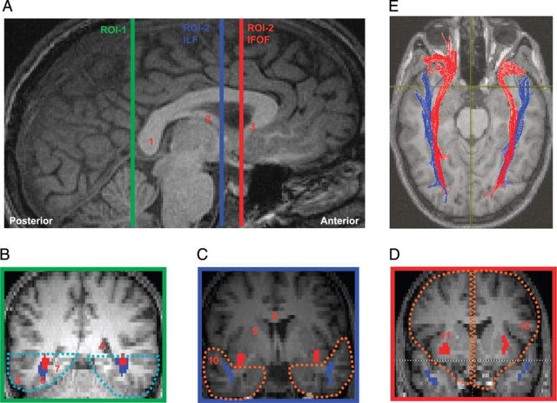
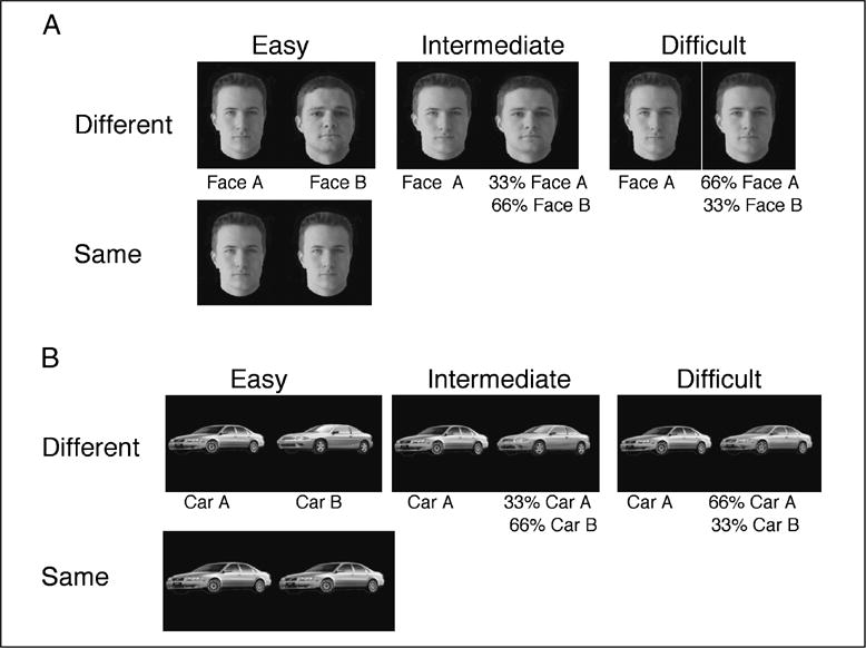
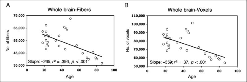
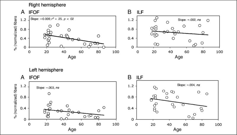
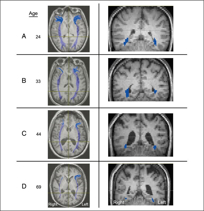
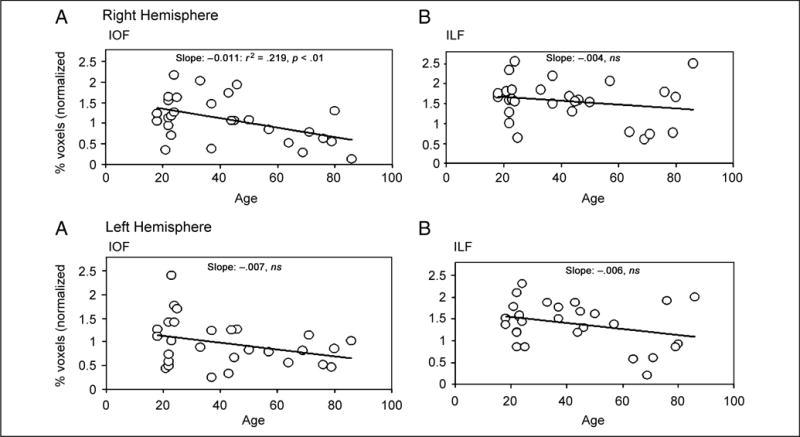
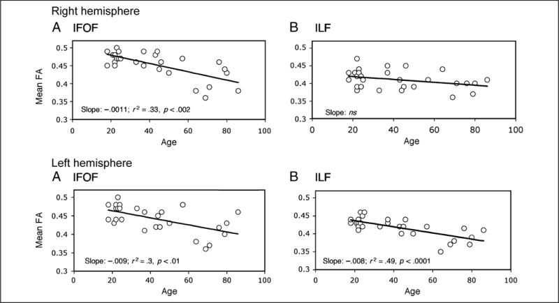
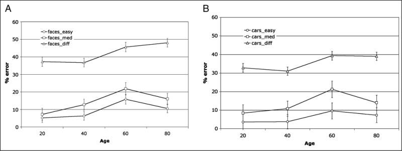
An age-related decline in face processing, even under conditions in which learning and memory are not implicated, has been well documented, but the mechanism underlying this perceptual alteration remains unknown. Here, we examine whether this behavioral change may be accounted for by a reduction in white matter connectivity with age. To this end, we acquired diffusion tensor imaging data from 28 individuals aged 18 to 86 years and quantified the number of fibers, voxels, and fractional anisotropy of the two major tracts that pass through the fusiform gyrus, the pre-eminent face processing region in the ventral temporal cortex. We also measured the ability of a subset of these individuals to make fine-grained discriminations between pairs of faces and between pairs of cars. There was a significant reduction in the structural integrity of the inferior fronto-occipital fasciculus (IFOF) in the right hemisphere as a function of age on all dependent measures and there were also some changes in the left hemisphere, albeit to a lesser extent. There was also a clear age-related decrement in accuracy of perceptual discrimination, especially for more challenging perceptual discriminations, and this held to a greater degree for faces than for cars. Of greatest relevance, there was a robust association between the reduction of IFOF integrity in the right hemisphere and the decline in face perception, suggesting that the alteration in structural connectivity between the right ventral temporal and frontal cortices may account for the age-related difficulties in face processing.


Figures

Figure 1
Figure 1
(A) Sagittal slice showing delineation of location for defining ROIs used for extracting the ILF and the IFOF. All ROIs were defined on each individual’s native space along the mid-sagittal plane. (B–D) The coronal slices are color coordinated to indicate their position on the mid-sagittal plane. (B) ROI-1, which encompasses the ventral occipito-temporal region is marked in cyan dotted lines in the coronal slice demarcated by the green line in (A). (C) ROI-2 for extracting ILF (blue fibers), which encompasses the anterior temporal lobe in each hemisphere, is marked in orange dotted lines in the coronal slice demarcated by the blue line in (A). Note that for extraction of the ILF fibers, the IFOF fibers (shown in red) were removed to avoid inclusion with the ILF fibers. (D) ROI-2 for extracting the IFOF (red fibers), which encompasses the frontal lobe in each hemisphere, is marked in orange dotted lines in the coronal slice demarcated by the red line in (A). Note that between the coronal slices corresponding to the blue and red lines marked on the sagittal plane in (A), the IFOF diverges toward the frontal cortex through the floor of the external capsule. 1 = Splenium of the callosum; 2 = Fornix; 3 = Rostrum of the callosum; 4 = Posterior horn of the lateral ventricle; 5 = Inferior temporal gyrus; 6 = Posterior fusiform gyrus; 7 = Inferior lingual gyrus; 8 = Body of the callosum; 9 = Internal capsule; 10 = Superior temporal gyrus; 11 = External capsule; 12 = Inferior frontal gyrus.
Figure 2
Figure 2
Examples of stimuli used in the (A) face and (B) car behavioral experiments. For each stimulus set, one quarter of the pairs of images were identical and required a “same” response, whereas the remaining three-quarters were different and required a “different” response. The different pairs could be easily discriminable (two entirely different faces or cars), intermediate in difficulty of discrimination (one face or car morphed with 66% of an entirely different image) or difficult to discriminate (one face or car morphed with an image containing 66% of itself and 33% of a different image).
Figure 3
Figure 3
Number of (A) fibers and (B) voxels through which fibers pass, calculated across the whole brain for each participant, plotted as a function of age. The slope, variance accounted for, and significance levels are shown for each graph.
Figure 4
Figure 4
The normalized percentage (as a function of whole brain) of fibers extracted from the right (upper panels) and left (lower panels) hemisphere for the IFOF (left) and ILF (right) as a function of age. As shown by the slope, it is only in the right IFOF where age accounts for a significant amount of the variance.
Figure 5
Figure 5
Representative axial (left) and coronal (right) slices taken from four participants, aged 24, 33, 44, and 69 years, revealing the loss of fibers as a function of increasing age, especially in the right hemisphere. Note that radiological convention is adopted with the right hemisphere depicted on the left of the image and vice versa.
Figure 6
Figure 6
The normalized percentage (as a function of whole brain) of voxels extracted from the right (upper panels) and left (lower panels) hemisphere for the IFOF (left) and ILF (right) as a function of age. As shown by the slope, it is only the right IFOF for which age accounts for a significant amount of the variance.
Figure 7
Figure 7
The fractional anisotropy (FA) values extracted from the right (upper panels) and left (lower panels) hemisphere for the IFOF (left) and ILF (right) as a function of age. As shown by the slope, age accounts for a significant amount of the variance for the right IFOF and left ILF and IFOF.
Figure 8
Figure 8
Mean percent error for performance on (A) faces and (B) car discrimination experiment for the different trials only (shown for easy, medium, and difficult trials) as a function of age. Note the three-way interaction between level of difficulty, age group, and stimulus type with the difficult face discrimination giving rise to more errors in the 60- and 80-year-olds than for any other condition or age group.

References

    1. Aharon I, Etcoff N, Ariely D, Chabris CF, O’Connor E, Breiter HC. Beautiful faces have variable reward value: fMRI and behavioral evidence. Neuron. 2001;32:537–551. - PubMed
    1. Alexander AL, Lobaugh NJ. Insights into brain connectivity using quantitative MRI measures of white matter. In: Jirasa V, McIntosh AR, editors. Handbook of brain connectivity. Berlin: Springer-Verlag; 2007. pp. 221–271.
    1. Avidan G, Hasson U, Malach R, Behrmann M. Detailed exploration of face-related processing in congenital prosopagnosia: 2. Functional neuroimaging findings. Journal of Cognitive Neuroscience. 2005;17:1150–1167. - PubMed
    1. Bar M, Kassam KS, Ghuman AS, Boshyan J, Schmid AM, Dale AM, et al. Top-down facilitation of visual recognition. Proceedings of the National Academy of Sciences, USA. 2006;103:449–454. - PMC - PubMed
    1. Bartzokis G, Beckson M, Lu PH, Nuechterlein KH, Edwards N, Mintz J. Age-related changes in frontal and temporal lobe volumes in men: A magnetic resonance imaging study. Archives of General Psychiatry. 2001;58:461–465. - PubMed

Publication types

MeSH terms