

Solution NMR structure of a designed metalloprotein and complementary molecular dynamics refinement
- PMID: 18275812
- PMCID: PMC3814030
- DOI: 10.1016/j.str.2007.11.011
Solution NMR structure of a designed metalloprotein and complementary molecular dynamics refinement
Abstract
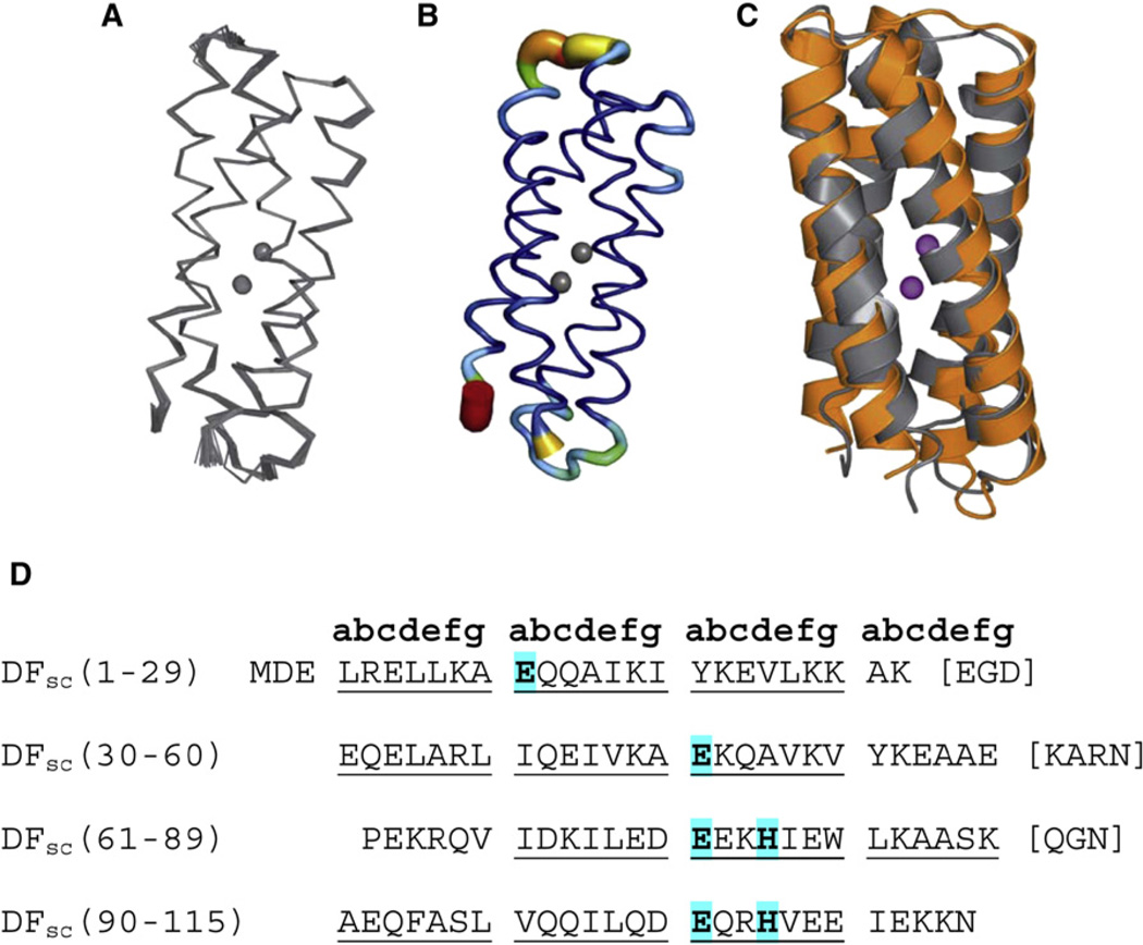
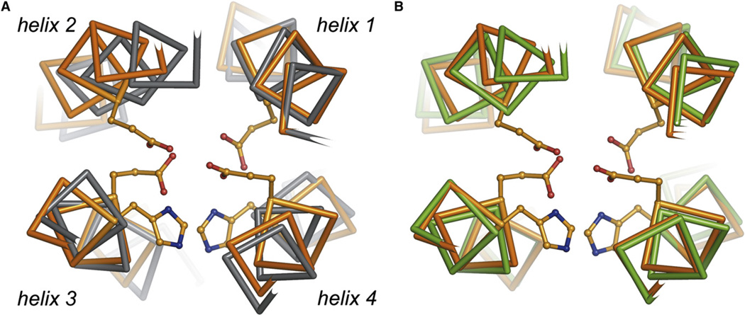
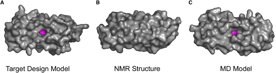
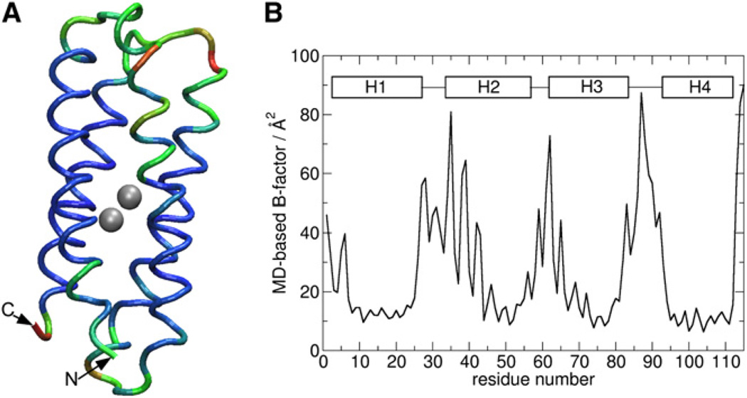
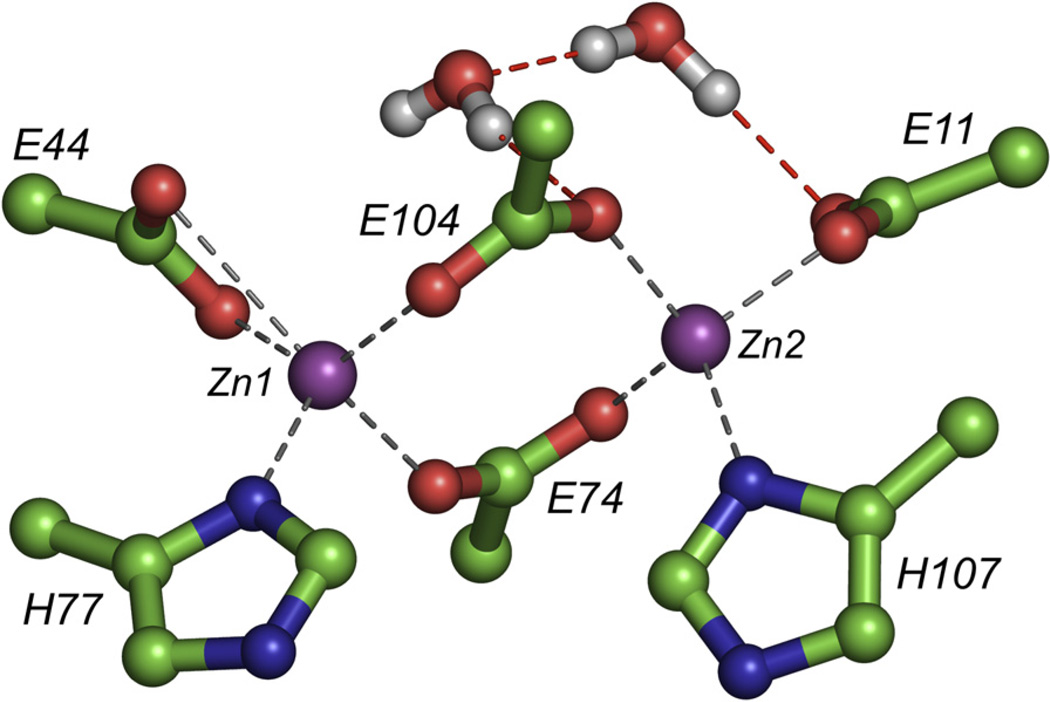
We report the solution NMR structure of a designed dimetal-binding protein, di-Zn(II) DFsc, along with a secondary refinement step employing molecular dynamics techniques. Calculation of the initial NMR structural ensemble by standard methods led to distortions in the metal-ligand geometries at the active site. Unrestrained molecular dynamics using a nonbonded force field for the metal shell, followed by quantum mechanical/molecular mechanical dynamics of DFsc, were used to relax local frustrations at the dimetal site that were apparent in the initial NMR structure and provide a more realistic description of the structure. The MD model is consistent with NMR restraints, and in good agreement with the structural and functional properties expected for DF proteins. This work demonstrates that NMR structures of metalloproteins can be further refined using classical and first-principles molecular dynamics methods in the presence of explicit solvent to provide otherwise unavailable insight into the geometry of the metal center.
Figures
References
-
- Bader RFW. Atoms in Molecules—A Quantum Theory. Volume 22. Oxford: Oxford University Press; 1990.
-
- Bansal M, Kumar S, Velavan R. HELANAL: a program to characterize helix geometry in proteins. J. Biomol. Struct. Dyn. 2000;17:811–819. - PubMed
-
- Becke AD. Density-functional exchange-energy approximation with correct asymptotic behavior. Phys. Rev. A. 1988;38:3098–3100. - PubMed
-
- Brunger AT, Adams PD, Clore GM, DeLano WL, Gros P, Grosse-Kunstleve RW, Jiang JS, Kuszewski J, Nilges M, Pannu NS, et al. Crystallography & NMR system: a new software suite for macromolecular structure determination. Acta Crystallogr. D Biol. Crystallogr. 1998;54:905–921. - PubMed
-
- Calhoun JR, Kono H, Lahr S, Wang W, DeGrado WF, Saven JG. Computational design and characterization of a monomeric helical dinuclear metalloprotein. J. Mol. Biol. 2003;334:1101–1115. - PubMed
Publication types
MeSH terms
Substances
Associated data
- Actions
Grants and funding
LinkOut - more resources
Full Text Sources
Miscellaneous
