. 2008 Apr 4;283(14):9239-47.
doi: 10.1074/jbc.M705175200. Epub 2008 Feb 13.

Focal adhesion kinase/Src suppresses early chondrogenesis: central role of CCN2

Affiliations

Focal adhesion kinase/Src suppresses early chondrogenesis: central role of CCN2

Daphne Pala et al. J Biol Chem. .

Abstract

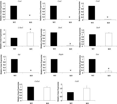
Adhesive signaling plays a key role in cellular differentiation, including in chondrogenesis. Herein, we probe the contribution to early chondrogenesis of two key modulators of adhesion, namely focal adhesion kinase (FAK)/Src and CCN2 (connective tissue growth factor, CTGF). We use the micromass model of chondrogenesis to show that FAK/Src signaling, which mediates cell/matrix attachment, suppresses early chondrogenesis, including the induction of Ccn2, Agc, and Sox6. The FAK/Src inhibitor PP2 elevates Ccn2, Agc, and Sox6 expression in wild-type mesenchymal cells in micromass culture, but not in cells lacking CCN2. Our results suggest a reduction in FAK/Src signaling is a critical feature permitting chondrogenic differentiation and that CCN2 operates downstream of this loss to promote chondrogenesis.


Figures

FIGURE 1.
FIGURE 1.
Loss of FAK in micromass culture increases chondrogenic differentiation. a, for protein analysis, Fak+/+ (WT) and -/- (KO) mouse mesenchymal cells (8.5 dpc) were plated in micromass cultures for 6 days. Protein isolated from mouse mesenchymal cells on day 6 was examined for FAK and CCN2 by Western blot analysis. FAK protein expression was absent in Fak-/- mouse embryo fibroblasts. CCN2 expression was up-regulated in the Fak-/- mouse embryo fibroblasts. β-actin was used as a loading control. b, PNA staining was performed on day 6. Fak-/- mouse embryo fibroblasts show markedly more condensation formations than Fak+/+ mouse embryo fibroblasts in the PNA-stained micromass cultures, as assessed by diameter of individual micromass cultures. Six different micromass cultures for each condition were examined. Representative cultures are shown. c, RNAs were harvested on day 6, and transcripts were analyzed by real-time RT-PCR. Ccn2, Col2a1, and L-Sox5 mRNA expression was significantly increased in Fak-/- mouse fibroblasts. Expression of Sox6 and Sox9 mRNAs did not change. Data shown represent means + S.E. from three independent experiments and are normalized to control GAPDH levels (each performed in triplicate). *, p < 0.05 using Student's paired t test. Note that Agc and Hapln mRNAs were not detected in any samples tested.
FIGURE 2.
FIGURE 2.
Loss of Ccn2 results in reduced mRNA expression of CCN family members and chondrogenic genes. Ccn2+/+ (WT) and -/- (KO) mouse mesenchymal cells were cultured for 48 h in monolayer, and gene expression was analyzed by real-time RT-PCR. Ccn2 mRNA expression was absent in Ccn2-/- mouse mesenchymal cells. Ccn1 and Ccn5 mRNA expression was significantly reduced in Ccn2-/- mouse mesenchymal cells. Moreover Sox6, Agc, Hapln, and Dcn mRNA was significantly reduced in Ccn2-/- mouse mesenchymal cells. Conversely, L-Sox5 mRNA expression was increased in Ccn2-/- mouse mesenchymal cells. Sox9, Col2a1, and ColX mRNA expression was not affected by loss of CCN2. Data shown represent means + S.E. relative to GAPDH control samples. Experiments were performed three times, with a variation <10%. *, p < 0.05 using Student's paired t test. Expression in WT cells was taken to represent 1.
FIGURE 3.
FIGURE 3.
In the absence of Ccn2, levels of early chondrogenic matrix-associated proteins Sox6 and aggrecan are decreased. A, protein isolated from Ccn2 +/+ (WT) and -/- (KO) mouse mesenchymal cells was cultured in monolayer for 48 h. Conditioned medium and protein extracts were examined for aggrecan and Sox6, respectively, by Western blot analysis. Both Sox6 and aggrecan protein expression were decreased in Ccn2-/- mouse mesenchymal cells. β-actin was used as a loading control. Densitometry analysis of aggrecan and Sox6, relative to β-actin, shows a significant difference between samples. No difference was seen in the expression of type II collagen and Sox5. Data shown represent means + S.E. from three trials. *, p <0.05 using Student's paired t test. B, for immunofluorescence analysis, cells were fixed in paraformaldehyde and stained with fluorescein isothiocyanate-labeled (FITC) antibody for Sox6 and with 4′,6-diamidino-2-phenylindole (DAPI) for nuclei. Sox6 was localized around the nuclei in the Ccn2 +/+ mouse mesenchymal cells, whereas expression was markedly decreased in the Ccn2-/- mouse mesenchymal cells. Six different fields were examined. A representative field is shown.
FIGURE 4.
FIGURE 4.
Loss of Ccn2 affects chondrogenesis in micromass culture. Ccn2+/+ (WT) and -/- (KO) mouse mesenchymal cells were subjected to micromass cultures for 6 days. PNA (a) and Alcian Blue (b) staining was performed on day 6. a, reduced condensation was observed in Ccn2-/- mouse mesenchymal cells, as assessed by micromass culture diameter. Six different micromass cultures for each condition were examined. Representative micromass cultures are shown. b, Ccn2+/+ cells show greater amount of Alcian Blue staining than the Ccn2-/- mouse mesenchymal cells. Staining of six different micromass cultures for each condition was examined. Staining was quantified as described under “Materials and Methods.” Average ± S.D. is shown. *, p < 0.05 using paired t test.
FIGURE 5.
FIGURE 5.
Loss of Ccn2 affects proteoglycan mRNA expression. Ccn2+/+ (WT) and -/- (KO) mouse mesenchymal cells were subjected to micromass cultures for 6 days. For real-time RT-PCR, RNA was harvested on days 1, 3, and 6. Agc, Sox6, and Hapln mRNA expression was significantly reduced in Ccn2-/- mouse mesenchymal cells on all 6 days examined. Conversely, Col2a1, L-Sox5, and Sox9 expression was significantly increased in Ccn2-/- mouse mesenchymal cells. Data shown are relative to GAPDH and represent means + S.E. from three independent experiments (each performed in triplicate). *, p < 0.05 using a two-way analysis of variance. Expression in WT cells on day 1 was taken to represent 1.
FIGURE 6.
FIGURE 6.
BMP-2 treatment of Ccn2-/- micromass cultures increases expression of chondrogenic extracellular matrix-associated genes. Ccn2+/+ (WT) and -/- (KO) mouse mesenchymal cells were subjected to micromass culture for 6 days. RNA was harvested on day 3, and transcripts were analyzed by real-time RT-PCR. a–c, induction of Agc, Sox6, and Col2a1 mRNA is greater in Ccn2-/- mouse mesenchymal cells compared with Ccn2+/+ mouse mesenchymal cells. Data shown are relative to GAPDH and represent means + S.E. from three independent experiments (each performed in triplicate). *, p < 0.05 using Student's paired t test. -Fold increase in response to BMP-2 is shown.
FIGURE 7.
FIGURE 7.
Inhibition of FAK/Src signaling increases mRNA expression of Ccn2 and chondrogenic matrix-associated genes. Ccn2+/+ (WT) and -/- (KO) mouse mesenchymal cells were subjected to micromass cultures. Each day for 6 days Me2SO or 10 μm FAK/Src inhibitor PP2 was added. RNA was harvested on day 6. mRNA levels of Ccn2, Agc, Hapln, and Sox6 were significantly increased compared with control cultures in Ccn2+/+ mouse mesenchymal cells treated with PP2, as determined by real-time RT-PCR. Such induction did not occur in Ccn2-/- mouse mesenchymal cells. Data shown are relative to GAPDH and represent means + S.E. from three independent experiments and are normalized to day 6 WT control sample and day 6 KO control sample (each performed in triplicate). *, p < 0.05 using a two-way analysis of variance. Note that we were not able to detect expression of CCN2 or Hapln RNA in KO cells.
FIGURE 8.
FIGURE 8.
Recombinant CCN2 induces Agc and Sox6 mRNA expression in Ccn2-/- mouse mesenchymal cells. Ccn2-/- (KO) mouse mesenchymal cells were cultured in monolayer and serum-starved for 24 h prior to treatment with or without full-length 38-kDa CCN2 (100 ng/ml) for 6 h. mRNA levels of Agc and Sox6 were significantly increased by treatment with CCN2. Hapln mRNA was undetectable in both untreated and treated Ccn2-/- mouse mesenchymal cells (not shown). Data shown are relative to GAPDH and represent means + S.E. from three independent experiments (each performed in triplicate). *, p < 0.05 using two-way analysis of variance. Expression in untreated KO sample was taken to represent 1.

Similar articles

Cited by

References

    1. Karsenty, G., and Wagner, E. F. (2002) Dev. Cell 2 389-406 - PubMed
    1. Kronenberg, H. M. (2003) Nature 423 332-336 - PubMed
    1. Zelzer, E., and Olsen, B. R. (2003) Nature 423 343-348 - PubMed
    1. Karsenty, G. (2003) Nature 423 316-318 - PubMed
    1. Cohn, M. J., and Tickle, C. (1996) Trends Genet. 12 253-257 - PubMed

Publication types

MeSH terms

LinkOut - more resources