. 2008 Mar;172(3):799-808.
doi: 10.2353/ajpath.2008.070918. Epub 2008 Feb 14.

Pathological expression of CXCL12 at the blood-brain barrier correlates with severity of multiple sclerosis

Affiliations

Pathological expression of CXCL12 at the blood-brain barrier correlates with severity of multiple sclerosis

Erin E McCandless et al. Am J Pathol. 2008 Mar.

Abstract

Dysregulation of blood-brain barrier (BBB) function and transendothelial migration of leukocytes are essential components of the development and propagation of active lesions in multiple sclerosis (MS). Animal studies indicate that polarized expression of the chemokine CXCL12 at the BBB prevents leukocyte extravasation into the central nervous system (CNS) and that disruption of CXCL12 polarity promotes entry of autoreactive leukocytes and inflammation. In the present study, we examined expression of CXCL12 and its receptor, CXCR4, within CNS tissues from MS and non-MS patients. Immunohistochemical analysis of CXCL12 expression at the BBB revealed basolateral localization in tissues derived from non-MS patients and at uninvolved sites in tissues from MS patients. In contrast, within active MS lesions, CXCL12 expression was redistributed toward vessel lumena and was associated with CXCR4 activation in infiltrating leukocytes, as revealed by phospho-CXCR4-specific antibodies. Quantitative assessment of CXCL12 expression by the CNS microvasculature established a positive correlation between CXCL12 redistribution, leukocyte infiltration, and severity of histological disease. These results suggest that CXCL12 normally functions to localize infiltrating leukocytes to perivascular spaces, preventing CNS parenchymal infiltration. In the patient cohort studied, altered patterns of CXCL12 expression at the BBB were specifically associated with MS, possibly facilitating trafficking of CXCR4-expressing mononuclear cells into and out of the perivascular space and leading to progression of disease.


Figures
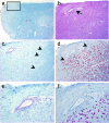
Figure 1
Figure 1
Active multiple sclerosis lesion within medulla. a: A section from the medulla of a postmortem specimen from a patient with MS stained with Luxol fast blue reveals multiple, irregularly bordered areas of demyelination. The boxed area indicates the region depicted at higher magnification after staining with H&E (b), Luxol fast blue (c), and ORO (d). Note the focus of demyelination with an irregular border (c, arrowheads), which includes a defined region bordered abruptly by ORO+ macrophages (d, arrowheads). The demyelinated region contains a venule with perivascular infiltration of small lymphocytes (e) that are adjacent to foamy ORO+ macrophages (f). Magnification: ×8 (a), ×40 (b–d), and ×100 (e and f).
Figure 2
Figure 2
CXCL12 redistribution occurs in venules within CNS tissues derived from MS patients. Endothelial cell localization (CD31, Alexa-488, green) of CXCL12 (Alexa-555, red) in arterioles and venules within postmortem CNS tissues derived from non-MS (a and b) and MS (c and d) patients. Note lack of CD31 staining within elastin layer of arteriole wall (large arrows). CXCL12 expression is detected along the basolateral (small arrow) and lumenal (arrowhead) surfaces of venules only in MS specimens (d). Nuclei are counterstained with ToPro3 (blue). Scale bar = 8 μm.
Figure 3
Figure 3
CXCL12 redistribution is specifically altered during MS. Endothelial cell localization (CD31, Alexa-488, green) of CXCL12 (Alexa-555, red) in cerebellar tissue obtained from non-MS (amyotropic lateral sclerosis) (a) and MS patients (b and c). Nuclei are counterstained with ToPro3 (blue). Scale bar = 10 μm. Quantification of fluorescence intensity aquired by confocal microscopy for CXCL12 (red stain and line) and CD31 (green stain and line) are shown for venules within non-MS (d) and MS (e and f) tissues. Double-headed arrows indicate location of line graph analysis. Three-dimensional reconstructions of microvessels stained with anti-CXCL12 (red) and anti-CD31 (green) antibodies are shown for venules depicted in a–c: non-MS (g), MS (h and i). The percentage of venules with loss of CXCL12 polarity in each patient in our non-MS and MS cohorts (j) were determined by examining CD31 and CXCL12 staining patterns in approximately 5 to 72 venules per patient (n = 6 control, 11 MS patients).
Figure 4
Figure 4
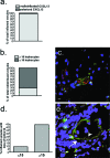
CXCL12 redistribution occurs in venules within active MS lesions regardless of extent of perivascular infiltrates. a: Combined analysis of all venules with ≥10 perivascular leukocytes within MS specimens were examined for redistributed CXCL12 expression (light gray bar) versus polarized expression (dark gray bar). b: Combined analysis of venules with CXCL12 redistribution for venules with ≥10 (light gray bar) versus ≤10 (dark gray bar) leukocytes adjacent to CD31+ endothelium. c: Endothelial cell localization (CD31, Alexa-488, green) of CXCL12 (Alexa-555, red) reveals loss of CXCL12 polarity in a venule without perivascular infiltrates (<10 associated leukocytes). d: Combined analysis of all venules with >15 versus <15 perivascular leukocytes for loss of perivascular CXCL12 expression. e: A CD31+ venule (Alexa-488, green) with intense perivascular infiltrates displays weak intralumenal (arrowhead) and no perivascular (arrow) expression of CXCL12. Scale bar = 8 μm.
Figure 5
Figure 5
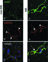
Astrocytes are a source of CXCL12. Activated astrocyte localization (GFAP, Alexa-488, green) (a) of CXCL12 (Alexa-555, red) (b, arrow) in the glial limitans adjacent to CXCL12-expressing endothelial cells (b, arrowheads) within a section from the optic nerve from a non-MS patient. c: Merged image. Scale bar = 8 μm. End-feet of activated astrocytes (GFAP, Alexa-488, green) (d) express increased staining for CXCL12 (Alexa-555, red) (e, arrowhead) within a section from the spinal cord from an MS patient. f: Merged image depicts colocalization of GFAP and CXCL12 (arrow).
Figure 6
Figure 6
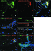
CXCR4 activation occurs within infiltrating leukocytes in active MS lesions. Cellular localization with panCXCR4 (Alexa-488, green) (a) and pS339-CXCR4 (Alexa-555, red) (b) antibodies in a section of the medulla with an active MS lesion. c: Note a subset of perivascular CXCR4-expressing cells (arrowheads) and endothelial cells (white arrow) contain activated CXCR4. Analyses of an inflamed CD31+ (Alexa-488, green) venule within sister-sections derived from an active MS lesion in the midbrain reveals redistribution of CXCL12 (Alexa-555, red) with decreased perivascular CXCL12 staining (d) is associated with activation of CXCR4 (Alexa-555, red) (f, arrowheads) within CD45+ leukocytes (Alexa-488, green) (e). The white line delineates the CD31+ venule perimeter (d, arrow) detected in the merged image (g). Analysis of CXCR4 activation in control CNS specimens reveals CD45+ (Alexa-488, green), pS339-CXCR4+ (Alexa-555, red) cells within the perivascular space in a thoracic cord section from a patient with CNS lymphoma (h) but no CXCR4 activation in CD45+ cells within the lumen of a vessel in the cerebrum of an amyotropic lateral sclerosis patient (i). Scale bar = 20 μm.
Figure 7
Figure 7
Redistribution of CXCL12 significantly correlates with histological markers of MS disease severity. Extents of inflammation (a), demyelination (b), and presence of macrophages (c) within CNS sections derived from MS patients were determined as described in the Materials and Methods, and severity scores versus percentage of CXCL12 redistribution for each block analyzed in the MS patient group are depicted. Correlation coefficient of best-fit line (r2) and P values are shown for each graph.

References

    1. Ballabh P, Braun A, Nedergaard M. The blood-brain barrier: an overview: structure, regulation, and clinical implications. Neurobiol Dis. 2004;16:1–13. - PubMed
    1. Frohman EM, Racke MK, Raine CS. Multiple sclerosis: the plaque and its pathogenesis. N Engl J Med. 2006;354:942–955. - PubMed
    1. Bielekova B, Goodwin B, Richert N, Cortese I, Kondo T, Afshar G, Gran B, Eaton J, Antel J, Frank JA, McFarland HF, Martin R. Encephalitogenic potential of the myelin basic protein peptide (amino acids 83–99) in multiple sclerosis: results of a phase II clinical trial with an altered peptide ligand. Nat Med. 2000;6:1167–1175. - PubMed
    1. Genain CP, Cannella B, Hauser SL, Raine CS. Identification of autoantibodies associated with myelin damage in multiple sclerosis. Nat Med. 1999;5:170–175. - PubMed
    1. Durán I, Martinez-Caceres EM, Rio J, Barbera N, Marzo ME, Montalban X. Immunological profile of patients with primary progressive multiple sclerosis: expression of adhesion molecules. Brain. 1999;122:2297–2307. - PubMed

Publication types