. 2008 Feb 15;319(5865):958-62.
doi: 10.1126/science.1147786.

A mouse model of mitochondrial disease reveals germline selection against severe mtDNA mutations

Affiliations

A mouse model of mitochondrial disease reveals germline selection against severe mtDNA mutations

Weiwei Fan et al. Science. .

Abstract

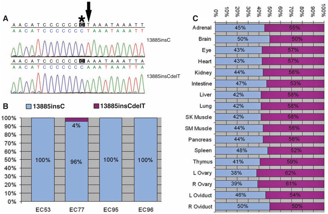
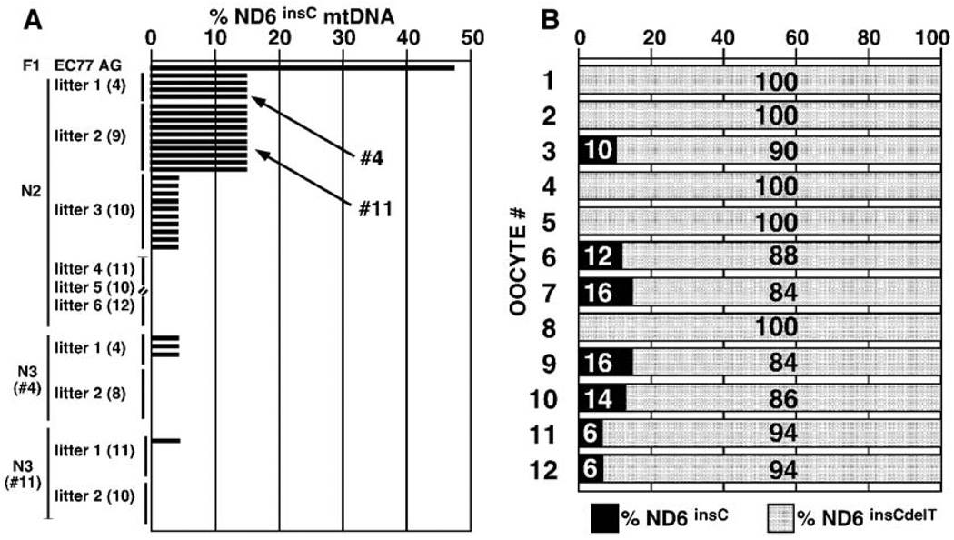
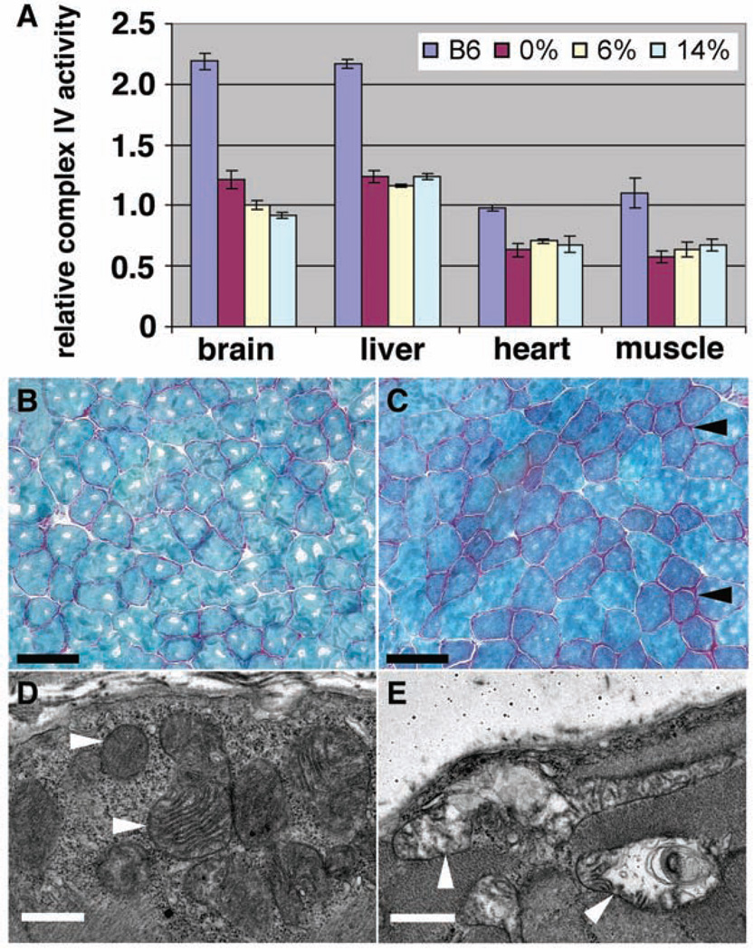
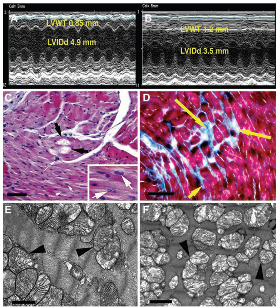
The majority of mitochondrial DNA (mtDNA) mutations that cause human disease are mild to moderately deleterious, yet many random mtDNA mutations would be expected to be severe. To determine the fate of the more severe mtDNA mutations, we introduced mtDNAs containing two mutations that affect oxidative phosphorylation into the female mouse germ line. The severe ND6 mutation was selectively eliminated during oogenesis within four generations, whereas the milder COI mutation was retained throughout multiple generations even though the offspring consistently developed mitochondrial myopathy and cardiomyopathy. Thus, severe mtDNA mutations appear to be selectively eliminated from the female germ line, thereby minimizing their impact on population fitness.


Figures

Fig. 1
Fig. 1
Qualitative and quantitative analysis of the ND6 frameshift (13885insC) and ND6 revertant (13885insCdelT) mutations in mouse ES cell cybrids and tissues of F1 female EC77-AG. (A) Sequence around nucleotide 13,885 of two clones of mtDNA from EC77 cells. Top (13885insC), the single C insertion causing the frameshift (asterisk); bottom (13885insCdelT), the T (arrow) deletion that restored the normal reading frame. (B) Percentages of ND6 frameshift (13885insC, blue) versus revertant (13885insCdelT, purple) in four independent mouse ES cybrids. (C) Proportions of ND6 frameshift (13885insC) and revertant (13885insCdelT) mtDNAs in the tissues of F1 female EC77-AG.
Fig. 2
Fig. 2
Selective elimination of ND6 frameshift mtDNA (13885insC) from F1 female EC77-AG and her offspring. (A) Percentages of ND6 frameshift (13885insC) mtDNAs in EC77-AG and her offspring, plus the pups of her daughters EC77 #4 and #11. Each offspring was analyzed from multiple litters. Litter sizes are indicated in parentheses. (B) Percentages of the ND6 frameshift (13885insC, black) and revertant (13885insCdelT, gray) mtDNAs in 12 oocytes isolated from EC77 progeny mice containing 14% of the ND6 frameshift mutation (13885insC).
Fig. 3
Fig. 3
Decreased mitochondrial complex IV activity and mitochondrial myopathy in COI mutant mice. (A) Complex IV activity was reduced to similar extents in various tissues of mutant mice harboring 0%, 6%, or 14% ND6 frameshift mutations plus 100% COI missense. Numbers of animals tested: six B6, seven 0% (0% ND6 frameshift plus 100% COI missense), three 6% (6% ND6 frameshift plus 100% COI missense), and four 14% (14% ND6 frameshift plus 100% COI missense), with three repeats performed for each test on each animal. (B and C) Gomori trichrome staining shows increased ragged red fibers (arrowheads) in skeletal muscle of 12-month-old COI mutant mice (C) compared to age-matched control (B). (D and E) Electron microscopy (EM) shows altered mitochondrial morphology (arrowheads) in skeletal muscle of 12-month-old COI mutant (E) mice compared to age-matched control (D). Scale bars, 75 µm [(B) and (C)], 1 µm [(D) and (E)].
Fig. 4
Fig. 4
Mitochondrial cardiomyopathy in 12-month-old mice homoplasmic for COI missense mutation. (A) Echocardiographic analysis of control heart. (B) Echocardiographic analysis of mutant heart showing increased left ventricular wall thickness (LVWT) and decreased left ventricular internal dimension in diastole (LVIDd). (C) Hematoxylin and eosin–stained mutant heart showing myofibrolysis (black arrows), myocyte hypertrophy (long white arrow), and binucleate cells (inset, white arrows). (D) Masson trichrome–stained mutant heart showing interstitial replacement fibrosis (yellow arrows). (E) EM of mitochondria (arrowheads) in normal heart. (F) EM of mutant heart showing mitochondrial proliferation, reduced matrix density, and cristolysis (arrowheads). Scale bars, 500 µm (C), 100 µm (D), 1 µm [(E) and (F)].

Comment in

References

    1. Wallace DC. Annu. Rev. Genet. 2005;39:359. - PMC - PubMed
    1. Wallace DC, Lott MT, Procaccio V. In: Emery and Rimoin’s Principles and Practice of Medical Genetics. ed. 5. Rimoin DL, Connor JM, Pyeritz RE, Korf BR, editors. vol. 1. Philadelphia: Churchill Livingstone; 2007. pp. 194–298.
    1. Schaefer AM, Taylor RW, Turnbull DM, Chinnery PF. Biochim. Biophys. Acta. 2004;1659:115. - PubMed
    1. Schaefer AM, et al. Ann. Neurol. 2007 10.1002/ana.21217.
    1. Bai Y, Attardi G. EMBO J. 1998;17:4848. - PMC - PubMed

Publication types

MeSH terms

Substances