. 2008 May 15;177(10):1111-21.
doi: 10.1164/rccm.200708-1243OC. Epub 2008 Feb 14.

Human rhinovirus 1B exposure induces phosphatidylinositol 3-kinase-dependent airway inflammation in mice

Affiliations

Human rhinovirus 1B exposure induces phosphatidylinositol 3-kinase-dependent airway inflammation in mice

Dawn C Newcomb et al. Am J Respir Crit Care Med. .

Abstract

Rationale: Infection with rhinovirus (RV) triggers exacerbations of asthma and chronic obstructive lung disease.

Objectives: We sought to develop a mouse model of RV employing RV1B, a minor group serotype that binds to the low-density lipoprotein receptor.

Methods: C57BL/6 mice were inoculated intranasally with RV1B, replication-deficient ultraviolet (UV)-irradiated RV1B, or RV39, a major group virus.

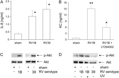
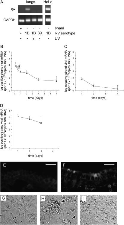
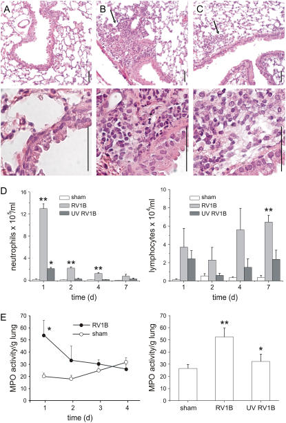
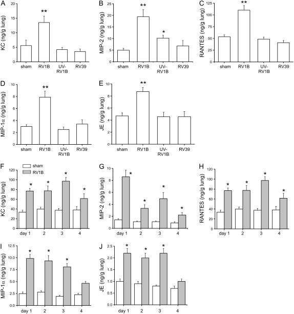
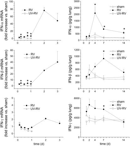
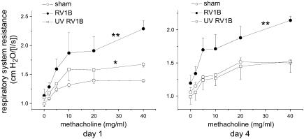
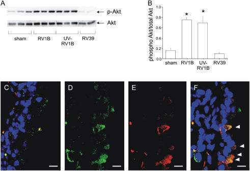
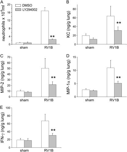
Measurements and main results: Viral RNA was present in the lungs of RV1B-treated mice, but not in those exposed to UV-irradiated RV1B or RV39. Lung homogenates of RV-treated mice contained infectious RV 4 days after inoculation. RV1B exposure induced neutrophilic and lymphocytic airway inflammation, as well as increased lung expression of KC, macrophage-inflammatory protein-2, and IFN-alpha and IFN-beta. RV1B-exposed mice showed airway hyperresponsiveness 1 and 4 days after inoculation. UV-irradiated RV1B induced modest neutrophilic airway inflammation and hyperresponsiveness 1 day after exposure. Both RV1B and UV-irradiated RV1B, but not RV39, increased lung phosphorylation of Akt. Confocal immunofluorescence showed colocalization of RV1B and phospho-Akt in the airway epithelium. Finally, pretreatment with the phosphatidylinositol 3-kinase inhibitor LY294002 attenuated chemokine production and neutrophil infiltration.

Conclusions: We conclude that RV1B induces airway inflammation in vivo. Evidence is presented that viral replication occurs in vivo and is required for maximal responses. On the other hand, viral replication was not required for a subset of RV-induced responses, including neutrophilic inflammation, airway hyperresponsiveness, and Akt phosphorylation. Finally, phosphatidylinositol 3-kinase/Akt signaling is required for maximal RV1B-induced airway neutrophilic inflammation, likely via its essential role in virus internalization.


Figures

<b>Figure 1.</b>
Figure 1.
Human rhinovirus 1B (RV1B) infection increases chemokine production and Akt phosphorylation in human and murine airway epithelial cells. (A) 16HBE14o human airway epithelial cells were infected with major group serotype RV39 or minor group serotype RV1B and conditioned medium was collected 48 hours postinfection and examined for IL-8/CXCL8 expression. (B) LA-4 murine airway epithelial cells were pretreated for 1 hour with LY294002 (10 μM) or dimethyl sulfoxide vehicle control and then infected with RV1B or mock-infected (sham) and murine IL-8 homolog, KC/CXCL1 was examined 48 hours postinfection. Bars represent the SEM for three experiments (*significantly different from sham, P < 0.05; **significantly different from sham and RV1B + LY294002, P < 0.05; one-way analysis of variance [ANOVA]). 16HBE14o (C) and LA-4 cells (D) were infected with RV1B or RV39 for 10 minutes and examined for phosphorylation of Akt. Immunoblots shown are typical of three separate experiments. UV = ultraviolet.
<b>Figure 2.</b>
Figure 2.
Viral RNA is detectable in the lungs of RV1B-treated mice. (A) Female C57BL/6 mice were inoculated with RV1B by intranasal instillation and lungs were examined by RT-PCR for viral RNA 1 day postinoculation. Mice were also exposed to ultraviolet (UV)-irradiated replication-deficient RV1B or RV39, a major group virus. As a positive control, HeLa cells were infected for 1 hour with RV1B at a multiplicity of infection of 10. RNA was extracted 16 hours postexposure and analyzed for viral RNA. Blots shown are typical of three separate experiments. GAPDH = glyceraldehyde-3-phosphate dehydrogenase. (B and C) Lungs of RV1B-inoculated mice were examined for positive-strand (B) and negative-strand (replicative) (C) rhinovirus (RV) RNA by quantitative PCR. Although positive-strand viral RNA decreased steadily after inoculation, viral RNA was detected up to 7 days postinoculation. There was a small but significant increase in positive-strand viral RNA copy number between the 12- and 18-hour time points (P = 0.043, one-way analysis of variance). We also detected a modest amount of negative-strand viral RNA, consistent with viral replication (n = 4–6, geometric mean ± SEM). (D) Positive-strand viral RNA was detected in the nasal washes of RV1B-inoculated mice up to 3 days after exposure (n = 3, geometric mean ± SEM). (E and F) One day postexposure, lungs from sham-inoculated (E) or RV1B-inoculated (F) mice were formalin-fixed and probed with purified RV1B antiserum. Specific staining for RV1B was noted in some but not all central airways. Scale bars: 20 μm. (GI) Supernatants from homogenized mouse lungs that were sham inoculated (G), inoculated with RV1B (H), or inoculated with UV-irradiated RV1B (I) were overlaid onto confluent HeLa cell monolayers and examined for cytopathic effect (arrowheads). Images shown are typical of three separate experiments (original magnification, ×100).
<b>Figure 3.</b>
Figure 3.
RV1B inoculation increases airway inflammation. Formalin-fixed, paraffin-embedded lungs harvested 1 day after viral exposure were stained with hematoxylin and eosin. (A) Sham-inoculated mice showed no inflammation. (B) RV1B-exposed mice demonstrated airway inflammation, indicated by arrows. (C) Mice exposed to ultraviolet (UV)-irradiated RV1B also showed evidence of inflammation (arrows). Scale bars: 50 μm. Images shown are typical of three separate experiments. (D) Bronchoalveolar lavage (BAL) was performed on mice 1 day postexposure and inflammatory cells were counted. Rhinovirus (RV)-exposed mice demonstrated increases in neutrophil (left) and lymphocyte (right) infiltration in the BAL fluid. (E) Left: Myeloperoxidase (MPO) activity was increased in RV1B-exposed mice 1 day postinoculation and declined to sham levels on Days 2 through 4. Right: In a separate experiment, mice exposed to UV-irradiated RV1B showed an intermediate increase in BAL neutrophil percentage and MPO activity. (Columns and error bars represent means ± SEM for three to nine mice. *Significantly different from sham, P < 0.05; **significantly different from sham and UV RV1B, P < 0.05; one-way analysis of variance.)
<b>Figure 4.</b>
Figure 4.
RV1B increases lung chemokine production. (AE) Mice were exposed to RV1B, replication-deficient ultraviolet (UV)-irradiated RV1B, or major group virus RV39, and chemokines were analyzed 1 day postexposure. KC/CXCL1, RANTES (regulated upon activation, T-cell expressed and secreted)/CCL5, macrophage-inflammatory protein (MIP)-1α/CCL3, and JE/CCL2 were increased with RV1B exposure, but not after inoculation with UV-irradiated RV1B or RV39. Mice inoculated with UV-irradiated RV1B showed an intermediate increase in MIP-2/CXCL2-3 expression. (FJ) Chemokine production was measured for 4 days after RV1B exposure. RV1B increased KC, MIP-2, RANTES, MIP-1α, and JE expression. (Error bars represent the SEM for 6–17 mice; *significantly different from sham, P < 0.05; **significantly different from sham and UV RV1B, P < 0.05; one-way analysis of variance.)
<b>Figure 5.</b>
Figure 5.
RV1B increases lung interferon production. Mice were exposed to HeLa cell lysate (sham inoculation), RV1B, or replication-deficient ultraviolet (UV)-irradiated RV1B, and interferon mRNA (left) and protein abundance (right) were analyzed 1–14 days postexposure. Relative to sham-inoculated mice, RV1B exposure increased IFN-α and IFN-β expression. (Data represent means ± SEM for three mice; *significantly different from sham inoculation and UV-irradiated RV, P < 0.05; one-way analysis of variance.)
<b>Figure 6.</b>
Figure 6.
RV1B infection increases airway cholinergic responsiveness. Mice were anesthetized and endotracheally intubated, and changes in respiratory system resistance to nebulized methacholine were measured with the flexiVent system (Scireq, Montreal, PQ, Canada). Mice were studied either 1 day (left) or 4 days (right) after viral exposure. Mice inoculated with RV1B, compared with sham-inoculated mice, demonstrated airway cholinergic responsiveness that was present 1 day after exposure and persisted to 4 days after exposure. One day after exposure, mice given ultraviolet (UV)-irradiated RV1B exhibited an intermediate state of airway responsiveness that was significantly increased compared with sham controls. (Data represent means ± SEM for three mice; *significantly different from sham inoculation, P < 0.05; **significantly different from sham and UV-irradiated RV1B; P < 0.05, two-way analysis of variance.)
<b>Figure 7.</b>
Figure 7.
A phosphatidylinositol 3-kinase downstream target, Akt, is activated in RV1B- and ultraviolet (UV)-RV1B–exposed mice. Whole lung homogenates collected 1 day postexposure were analyzed for Ser473 Akt phosphorylation and total Akt expression. (A) Exposure to RV1B and UV-irradiated RV1B each increased Akt phosphorylation compared with sham inoculation (representative blot shown). (B) Densitometry showing RV1B- or UV RV1B–induced increases in Akt phosphorylation when compared with total Akt. Error bars represent the SEM for six mice (*significantly different compared with sham and RV39, P < 0.05; one-way analysis of variance). (CF) RV1B and phospho-Akt colocalize in airway epithelial cells. Confocal fluorescence images demonstrate staining for RV1B (green), phospho-Akt (red), and airway nuclei (blue). For each image, the airway lumen is oriented to the right, and scale bars represent a length of 10 μm. (C) A merged image from a large airway of a sham-inoculated mouse. There is no specific staining of the airway epithelium. (D), (E), and (F) show green, red, and merged images, respectively, of a large airway from an RV1B-exposed mouse. Note orange-appearing colocalization of RV1B and phosphorylated Akt in four individual airway epithelial cells (arrowheads). Images shown are typical of three separate experiments.
<b>Figure 8.</b>
Figure 8.
Phosphatidylinositol 3-kinase is required for maximal RV1B-induced chemokine production in vivo. Mice were pretreated with LY294002 (3 mg/kg body weight) or vehicle (dimethyl sulfoxide [DMSO]) and exposed to RV1B or sham treatment 1 hour later. (A) One day postexposure, LY294002 decreased the bronchoalveolar lavage neutrophil percentage. (BE) LY294002 attenuated RV1B-induced KC, MIP-2, MIP-1α, and IFN-γ expression, respectively. (Error bars represent the SEM for eight or nine mice; **significantly different compared with RV1B and DMSO, P < 0.05; one-way analysis of variance.)

References

    1. Nicholson KG, Kent J, Ireland DC. Respiratory viruses and exacerbations of asthma in adults. BMJ 1993;307:982–986. - PMC - PubMed
    1. Kling S, Donninger H, Williams Z, Vermeulen J, Weinberg E, Latiff K, Ghildyal R, Bardin P. Persistence of rhinovirus RNA after asthma exacerbation in children. Clin Exp Allergy 2005;35:672–678. - PubMed
    1. Greenberg SB, Allen M, Wilson J, Atmar RL. Respiratory viral infections in adults with and without chronic obstructive pulmonary disease. Am J Respir Crit Care Med 2000;162:167–173. - PubMed
    1. Seemungal T, Harper-Owen R, Bhowmik A, Moric I, Sanderson G, Message S, Maccallum P, Meade TW, Jeffries DJ, Johnston SL, et al. Respiratory viruses, symptoms, and inflammatory markers in acute exacerbations and stable chronic obstructive pulmonary disease. Am J Respir Crit Care Med 2001;164:1618–1623. - PubMed
    1. Papadopoulos NG, Bates PJ, Bardin PG, Papi A, Leir SH, Fraenkel DJ, Meyer J, Lackie PM, Sanderson G, Holgate ST, et al. Rhinoviruses infect the lower airways. J Infect Dis 2000;181:1875–1884. - PubMed

Publication types

MeSH terms