

doi: 10.1038/nchembio0308-148.
A place for thioether chemistry in cellular copper ion recognition and trafficking
Affiliations
- PMID: 18277969
- PMCID: PMC2265432
- DOI: 10.1038/nchembio0308-148
Item in Clipboard
A place for thioether chemistry in cellular copper ion recognition and trafficking
Nat Chem Biol.
2008 Mar.
Abstract
Over the last decade, cysteine thiolate ligands have been shown to be critical to the Cu(I) (cuprous) binding chemistry of many cytosolic metallochaperone and metalloregulatory proteins involved in copper physiology. More recently, the thioether group of methionine has begun to emerge as an important Cu(I) ligand for trafficking proteins in more oxidizing cellular environments.
Figures
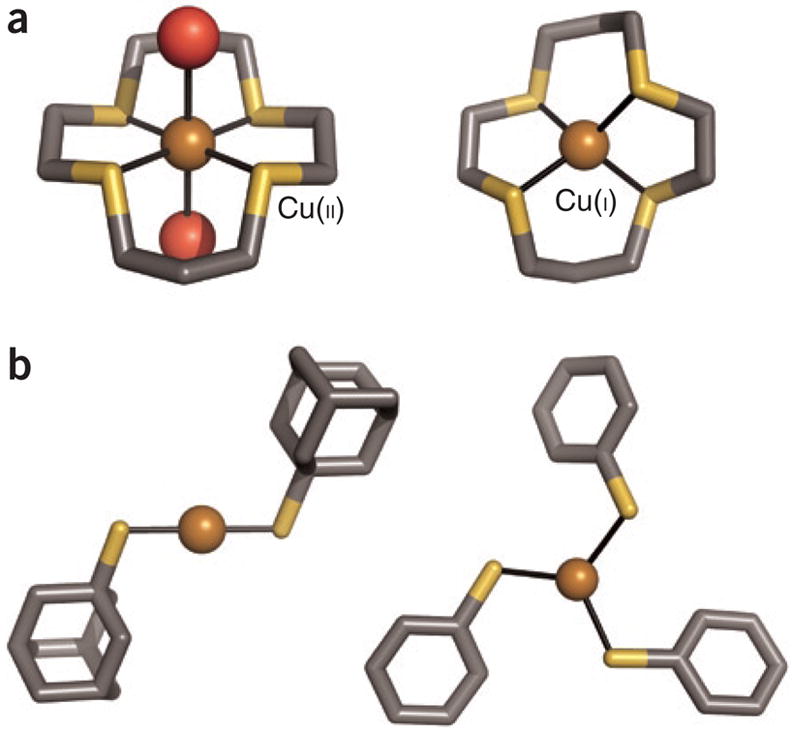
Four crystallographically characterized small-molecule complexes are shown (gray, C; dark yellow, S; red, O; bronze, Cu), labeled with the copper oxidation state. (a) The Cu(II)–thioether macrocycle complex, left, shows a higher coordination number (with two water ligands) than that of the reduced Cu(I) complex (ref. and references therein). (The macrocycle structure has been simplified for the figure.) (b) Discrete two-and three-coordinate Cu(I)-thiolate complexes have been obtained with the use of sterically encumbering ligands and appropriate counterions,. The discussionof Cu(I) coordination chemistry is simplified here, with certain nitrogen-donor ligands, such as the phenanthroline-based bathocuproine disulfonic acid (BCS), acting as very tight Cu(I) complexing agents.
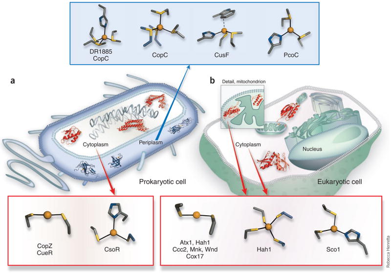
Characterized Cu(I) trafficking and sensing coordination sites classified by cellular localization and function. (a) Protein structures depicted in theprokaryotic cell are the periplasmic putative trafficking proteins, which all form positively charged Cu(I) complexes (colored blue): Cu(I)-DR1885 (ref. 25),Cu(I)-CusF26 and Cu(I)2CopC224); and the cytoplasmic proteins, which all form negatively charged Cu(I) complexes (colored red): the chaperone Cu(I)-CopZ11and the metalloregulatory DNA-binding proteins Cu(I)2CsoR2 (ref. 15) and Cu(I)2CueR2 (ref. 6). (b) In the cytosol of the eukaryotic cell (not drawn to scale),two chaperone structures are shown, the yeast Cu(I)-Atx1 (ref. 7) and the human Cu(I)-Hah12 (ref. 12). A number of mitochondrial Cco assembly proteinshave suspected Cu(I) transport function, and the Cu(I)-Sco1 (ref. 17) and Cu(I)-Cox17 (ref. 20) structures are shown. The coordination sites are shown indetail outside of the cell diagrams, and have been characterized by a combination of crystallographic, XAS and NMR structural studies. In addition to theprotein sites listed above, a Met3His site of CopC23 and a Met2His site of PcoC22 are included with the methionine-rich cationic sites of the prokaryoticperiplasmic proteins. The anionic Cys2Cu(I) sites of the eukaryotic cytosolic proteins Hah1 (ref. 30) and Atx1 (ref. 7) and the cytosolic metal-bindingdomains of the Ccc2, Menkes (Mnk)9 and Wilson (Wnd)10 disease Cu(I) transporting P-type ATPases are also included.
References
-
- Finney LA, O’Halloran TV. Science. 2003;300:931–936. - PubMed
-
- Rorabacher DB. Chem Rev. 2004;104:651–697. - PubMed
-
- Zeevi S, Tshuva EY. Eur J Inorg Chem. 2007:5369–5376.
-
- Schafer FQ, Buettner GR. Free Radic Biol Med. 2001;30:1191–1212. - PubMed
-
- Rae TD, Schmidt PJ, Pufahl RA, Culotta VC, O'Halloran TV. Science. 1999;284:805–808. - PubMed
Publication types
MeSH terms
Substances
Grants and funding
LinkOut - more resources
Full Text Sources
Other Literature Sources
