Review
. 2008 Mar;4(3):186-93.
doi: 10.1038/nchembio.71.

Versatility of biological non-heme Fe(II) centers in oxygen activation reactions

Affiliations
Review

Versatility of biological non-heme Fe(II) centers in oxygen activation reactions

Elena G Kovaleva et al. Nat Chem Biol. 2008 Mar.

Abstract

Oxidase and oxygenase enzymes allow the use of relatively unreactive O2 in biochemical reactions. Many of the mechanistic strategies used in nature for this key reaction are represented within the 2-histidine-1-carboxylate facial triad family of non-heme Fe(II)-containing enzymes. The open face of the metal coordination sphere opposite the three endogenous ligands participates directly in the reaction chemistry. Here, data from several studies are presented showing that reductive O2 activation within this family is initiated by substrate (and in some cases cosubstrate or cofactor) binding, which then allows coordination of O2 to the metal. From this starting point, the O2 activation process and the reactions with substrates diverge broadly. The reactive species formed in these reactions have been proposed to encompass four oxidation states of iron and all forms of reduced O2 as well as several of the reactive oxygen species that derive from O-O bond cleavage.


Figures
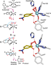
Figure 1
Figure 1
Oxygen activation by extradiol dioxygenases. This type of activation involves simultaneous activation of O2 and substrate, once both are bound to the active site Fe(II). It proceeds through formation of an alkylperoxo intermediate in which activated dioxygen attacks the substrate before O–O bond cleavage. The R-groups of the substrates considered here are –CH2–COO (HPCA, 1) or –NO2 (4NC, 2). The structures shown are the Brevibacterium fuscum HPCD 4NC-semiquinone–Fe(II)–O2•− (top) and Fe(II)- alkylperoxo (bottom) intermediates (PDB 2IGA) Atom color code: gray, carbon (enzyme residues); yellow, carbon (substrate); blue, nitrogen; red, oxygen; cyan, iron. Red and grey dashed lines show hydrogen bonds and potential bonds to iron, respectively.
Figure 2
Figure 2
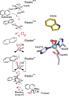
Oxygen activation by Rieske cis-diol dioxygenases. This type of reaction requires formation of a more reactive species that is found in the extradiol dioxygenase pathway of Fig. 1. One electron from the Rieske cluster and one from the Fe(II) are used to form Fe(III)–OOH which may then form Fe(V)=O–OH as the reactive species. The structure shown is of the hydroperoxo intermediate of Pseudomonas sp. strain NCIB 9816-4 NDO with the substrate analog indole bound nearby (PDB 1O7N) Atom color code: gray, carbon (enzyme residues); yellow, carbon (substrate); blue, nitrogen; red, oxygen; cyan, iron.
Figure 3
Figure 3
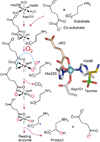
Oxygen activation by 2-oxo-acid dioxygenases. An initial Fe(III)–O2•− species attacks the Fe-bound αKG co-substrate to yield an Fe(IV)=O reactive species. This species, in turn, attacks the substrate by hydrogen atom abstraction. The structure shown is of the initial reactive complex of Escherichia coli TauD with αKG bound to the iron and taurine bound nearby (PDB 1GQW) Atom color code: gray, carbon (enzyme residues); yellow, carbon (substrate); bronze, carbon (co-substrate); blue, nitrogen; red, oxygen; green, sulfur; cyan, iron.
Figure 4
Figure 4
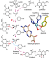
Oxygen activation by tetrahydropterin-dependent hydroxylases. A peroxo bridge is formed between Fe(II) and an electron donating tetrahydropterin cofactor in this O2 activation mechanism. The stability of the 4a-peroxypterin adduct allows the heterolytic O–O bond cleavage to yield an Fe(IV)=O species that reacts, in turn, with the aromatic substrate by electrophilic aromatic substitution. The structure shown is of human phenylalanine hydroxylase complexed with a substrate analog and tetrahydropterin (PDB 1MMK) . Atom color code: gray, carbon (enzyme residues); yellow, carbon (substrate); bronze, carbon (co-substrate); blue, nitrogen; red, oxygen; green, sulfur; cyan, iron.
Figure 5
Figure 5
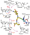
Oxygen activation in the mechanism of formation of isopenicillin N by IPNS. Binding of O2 to the active site Fe(II) yields an ACV-bound Fe(III)–O2•− reactive intermediate. Hydrogens (orange) and electrons derived from the closure of the β-lactam ring are then used to break the O–O bond and generate an Fe(IV)=O intermediate that is proposed to serve as a regent to effect the thiazolidine ring closure reaction. The structure shown is of Aspergillus nidulans IPNS complexed with ACV (PDB 1BK0) Atom color code: gray, carbon (enzyme residues); yellow, carbon (substrate); blue, nitrogen; red, oxygen; green, sulfur; cyan, iron.
Figure 6
Figure 6
Oxygen activation by the flexible 2-His+Asp/Glu facial triad motif. All reactions begin by binding a substrate or cofactor to or near the iron resulting in release of solvents to open an O2 binding site on the iron. A myriad of downstream chemistries are possible as determined by the nature of the exogenous molecules and active site residues near the catalytic surface of the iron. The three ligand sites shown to be occupied by H2O in the central structure can be vacant, occupied by OH,or occupied by a weak protein ligand in different enzymes from the 2-His+Asp/Glu family.

References

    1. Ozer A, Bruick RK. Non-heme dioxygenases: cellular sensors and regulators jelly rolled into one? Nat. Chem. Biol. 2007;3:144–153. - PubMed
    1. Kirk TK. In: Degradation of lignin. in Microbial Degradation of Organic Compounds. Gibson DT, editor. Vol. 13. New York, New York: Marcel Dekker, Inc; 1984. pp. 399–438.
    1. Hakemian AS, Rosenzweig AC. The Biochemistry of Methane Oxidation. Annu. Rev. Biochem. 2007;76:223–241. - PubMed
    1. Gibson DT, Parales RE. Aromatic hydrocarbon dioxygenases in environmental biotechnology. Curr. Opin. Biotechnol. 2000;11:236–243. - PubMed
    1. Baldwin JE, Abraham E. Biosynthesis of penicillins and cephalosporins. Nat. Prod. Rep. 1988;5:129–145. - PubMed

Publication types