. 2008 Apr 15;228(2):190-9.
doi: 10.1016/j.taap.2007.12.015. Epub 2007 Dec 23.

Nickel compounds induce histone ubiquitination by inhibiting histone deubiquitinating enzyme activity

Affiliations

Nickel compounds induce histone ubiquitination by inhibiting histone deubiquitinating enzyme activity

Qingdong Ke et al. Toxicol Appl Pharmacol. .

Abstract

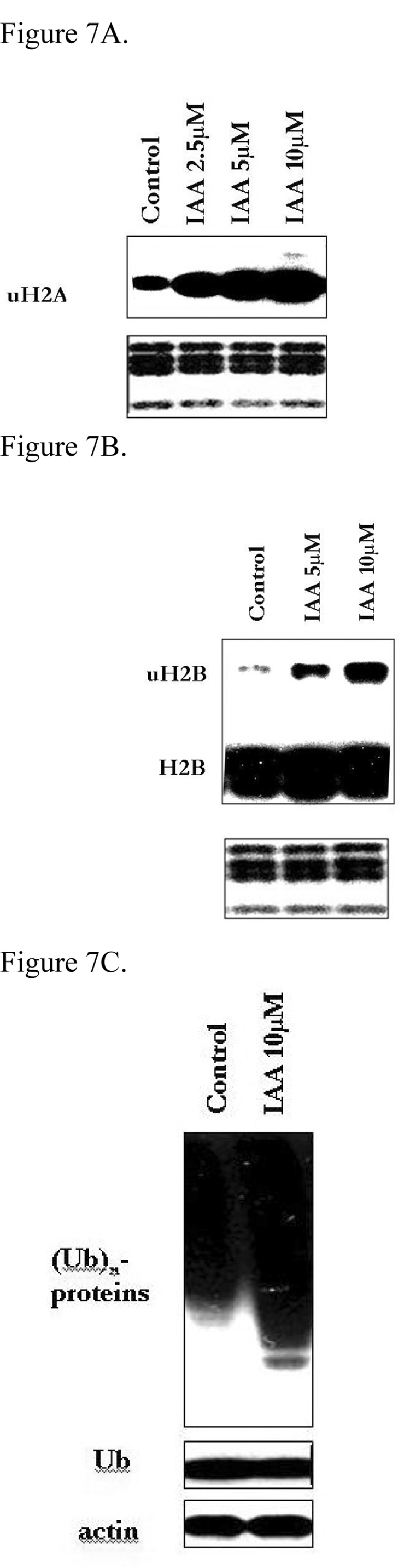
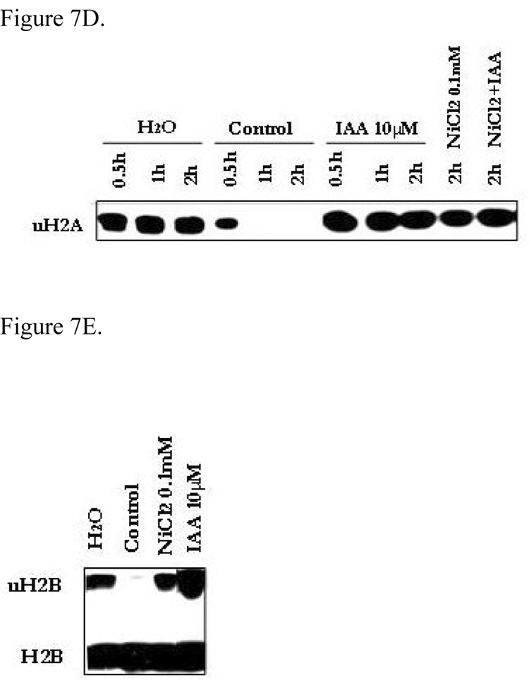
Nickel (Ni) compounds are known carcinogens but underlying mechanisms are not clear. Epigenetic changes are likely to play an important role in nickel ion carcinogenesis. Previous studies have shown epigenetic effects of nickel ions, including the loss of histone acetylation and a pronounced increase in dimethylated H3K9 in nickel-exposed cells. In this study, we demonstrated that both water-soluble and insoluble nickel compounds induce histone ubiquitination (uH2A and uH2B) in a variety of cell lines. Investigations of the mechanism by which nickel increases histone ubiquitination in cells reveal that nickel does not affect cellular levels of the substrates of this modification, i.e., ubiquitin, histones, and other non-histone ubiquitinated proteins. In vitro ubiquitination and deubiquitination assays have been developed to further investigate possible effects of nickel on enzymes responsible for histone ubiquitination. Results from the in vitro assays demonstrate that the presence of nickel did not affect the levels of ubiquitinated histones in the ubiquitinating assay. Instead, the addition of nickel significantly prevents loss of uH2A and uH2B in the deubiquitinating assay, suggesting that nickel-induced histone ubiquitination is the result of inhibition of (a) putative deubiquitinating enzyme(s). Additional supporting evidence comes from the comparison of the response to nickel ions with a known deubiquitinating enzyme inhibitor, iodoacetamide (IAA). This study is the first to demonstrate such effects of nickel ions on histone ubiquitination. It also sheds light on the possible mechanisms involved in altering the steady state of this modification. The study provides further evidence that supports the notion that nickel ions alter epigenetic homeostasis in cells, which may lead to altered programs of gene expression and carcinogenesis.


Figures

Figure 1
Figure 1. Nickel compounds induce ubiquitination of histone H2A and H2B in cells
(A) The effects of NiCl2 and Ni3S2 on cell proliferation in A549 cells. A549 cells were seeded one day before the treatment and treated with 0.1, 0.5, and 1 mM NiCl2, or 0.5, 1.0, and 2.5 µg/cm2 Ni3S2 for 24 hr or 48 hr. At the end of exposure, the cells were washed with 1x PBS, trypsinized and counted. The cell number from each treatment was compared with that from the untreated control. (B) A549 cells were treated with 0.25, 0.5, and 1.0 mM NiCl2 or 0.5 and 1.0 µg/cm2 Ni3S2 for 24 hr. Isolated histones (5 µg/treatment for detection of uH2A and 15 µg/treatment for detection of uH2B) were separated over 15% SDS-PAGE gels and subjected to Western blotting with the antibody against uH2A or H2B. The lower panels show the gels stained with Coomassie Blue after transfer to monitor for histone loading. (C) A549 cells were treated with 1.0 mM NiCl2 for 4, 8, 12, 24, 48, and 72 hr. (D) Cl41, Beas-2B, HeLa, and Hep3B cells were treated with 0.5 or 1.0 mM NiCl2 for 24 hr. (E) A549 cells were treated with 0.25, 0.5, and 1.0 mM NiCl2 or 0.5 and 1.0 µg/cm2 Ni3S2 for 24 hr. (F) Cl41 cells were treated with 0.5 or 1.0 mM NiCl2 for 24 hr.
Figure 2
Figure 2. Schematic of ubquitination and deubiquitination of histone H2A and H2B by ubiquitinating enzymes (E1, E2, and E3) and deubiquitinating enzymes, respectively
Figure 3
Figure 3. Nickel does not affect the cellular levels of ubiquitin, H2A, or H2B
A549 cells were treated with 0.25, 0.5, and 1.0 mM NiCl2 for 24 hr. (A) Whole protein extracts (40 µg) were separated over 15% SDS-PAGE gels and subjected to Western blotting with antibody against ubiquitin (Ub). The same membrane was reblotted with the antibody against actin (loading control). (B) Isolated total histones (5 µg) were separated over 15% SDS-PAGE gels and the gels were stained with Coomassie Blue.
Figure 4
Figure 4. Nickel does not affect the levels of total non-histone ubiquitinated proteins in cells
(A) A549 cells were treated with 1.0 mM NiCl2 for 24 hr. Whole protein extracts (40 µg) were separated over 4–15% SDS-PAGE gels and subjected to Western blotting with antibody against Ub. The same membrane was reblotted with the antibody against actin (loading control). (B) A549 cells were treated with 5 µM MG132, or 1.0 mM NiCl2 for 2, 4, 8, 12, and 24 hr. Whole protein extracts (40 µg) were separated over 4–15% SDS-PAGE gels and subjected to Western blotting with antibody against Ub. The same membrane was reblotted with the antibody against actin (loading control). Isolated histones (5 µg) were separated over 15% SDS-PAGE gels and subjected to Western blotting with antibody against uH2A. The lower panels show the gels stained with Coomassie Blue after transfer in order to monitor for histone loading.
Figure 5
Figure 5. Nickel does not enhance the level of uH2A in the in vitro ubiquitination assay
(A) Schematic of the in vitro ubiquitination assay. (B) Histones (5 µg) were incubated with ubiquitination mix containing ubiquitin (1 µg), cell lysate (20 µg) from A549 cells, and 0.5 mM ATP, in the absence or presence of NiCl2 (0.1 and 1 mM). After incubation at 37°C for 1 hr, the reaction was terminated by addition of SDS-PAGE loading buffer and subjected to Western blot with the antibody against uH2A. The lower panels show the same membrane reblotted with the antibody against ubiquitin.
Figure 6
Figure 6. Nickel prevents the loss of uH2A in the in vitro deubiquitination assay
(A) Schematic of the in vitro ubiquitination assay. (B) Total histones (5 µg) extracted from A549 cells were incubated with deubiquitination mix containing 20 µg of cell lysate from A549 cells, in the absence or presence of NiCl2 (0.1, 1, and 10 mM). Without incubation or after incubation at 37°C for 1 hr, the reaction was terminated by addition of SDS-PAGE loading buffer and subjected to Western blot with the antibody against uH2A. The same blots were reblotted with the antibody against ubiquitin (Ub). (C) Histones were incubated with 20 µg of the cell lysate in the absence or presence of NiCl2 (0.1 mM). After incubation at 37 °C for 0.5, 1, 2, or 4 hr, the reaction was terminated and samples processed as above.
Figure 7
Figure 7. Iodoacetamide (IAA) - a deubiquitinating enzyme inhibitor – induces ubiquitinated histones in cells and prevents the loss of ubiquitinated histone in vitro
(A) A549 cells were treated with IAA (2.5, 5, or 10 µM) for 24 hr. Isolated histones (5 µg) were separated over 15% SDS-PAGE gels and subjected to Western blotting with the antibody against uH2A. The lower panels show the gels stained with Coomassie Blue after transfer to monitor for histone loading. (B) A549 cells were treated with IAA (5 or 10 µM) for 24 hr. Isolated histones (15 µg) were separated over 15% SDS-PAGE gels and subjected to Western blot with the antibody against uH2B. The lower panels show the gels stained with Coomassie Blue after transfer to monitor for histone loading. (C) A549 cells were treated with 10 µM IAA for 24 hr. Whole protein extracts (40 µg) were separated over 4–15% SDS-PAGE gels and subjected to Western blotting with antibody against Ub. The same membrane was reblotted with the antibody against actin (loading control). (D) Total histones (5 µg) were incubated with 20 µg of protein extracts from A549 cells in the absence or presence of IAA (10 µM) and/or NiCl2 (0.1 mM). After incubation at 37 °C for 0.5, 1, and 2 hr, the reaction was terminated by addition of SDS-PAGE loading buffer and subjected to Western blot with the antibody against uH2A. (E) Total histones (5 µg) were incubated with 20 µg of protein extracts from A549 cells in the absence or presence of IAA (10 µM) or NiCl2 (0.1 mM). After incubation at 37 °C for 1 hr, the reaction was terminated by addition of SDS-PAGE loading buffer and subjected to Western blot with the antibody against H2B.
Figure 8
Figure 8. Model of the mechanism by which nickel induces histone ubiquitination
Figure 9
Figure 9. Model of the epigenetic effects of nickel compounds

References

    1. Biedermann KA, Landolph JR. Induction of anchorage independence in human diploid foreskin fibroblasts by carcinogenic metal salts. Cancer Res. 1987;47:3815–3823. - PubMed
    1. Biggart NW, Costa M. Assessment of the uptake and mutagenicity of nickel chloride in salmonella tester strains. Mutat Res. 1986;175:209–215. - PubMed
    1. Broday L, Cai J, Costa M. Nickel enhances telomeric silencing in Saccharomyces cerevisiae. Mutat Res. 1999;440:121–130. - PubMed
    1. Cao R, Tsukada Y, Zhang Y. Role of Bmi-1 and Ring1A in H2A ubiquitylation and Hox gene silencing. Mol Cell. 2005;20:845–854. - PubMed
    1. Chen H, Ke Q, Kluz T, Yan Y, Costa M. Nickel ions increase histone H3 lysine 9 dimethylation and induce transgene silencing. Mol Cell Biol. 2006;26:3728–3737. - PMC - PubMed

Publication types