. 2008 Mar 31;411(1-2):46-58.
doi: 10.1016/j.gene.2008.01.003. Epub 2008 Jan 17.

Molecular and functional characterization of a tandem-repeat galectin from the freshwater snail Biomphalaria glabrata, intermediate host of the human blood fluke Schistosoma mansoni

Affiliations

Molecular and functional characterization of a tandem-repeat galectin from the freshwater snail Biomphalaria glabrata, intermediate host of the human blood fluke Schistosoma mansoni

Timothy P Yoshino et al. Gene. .

Abstract

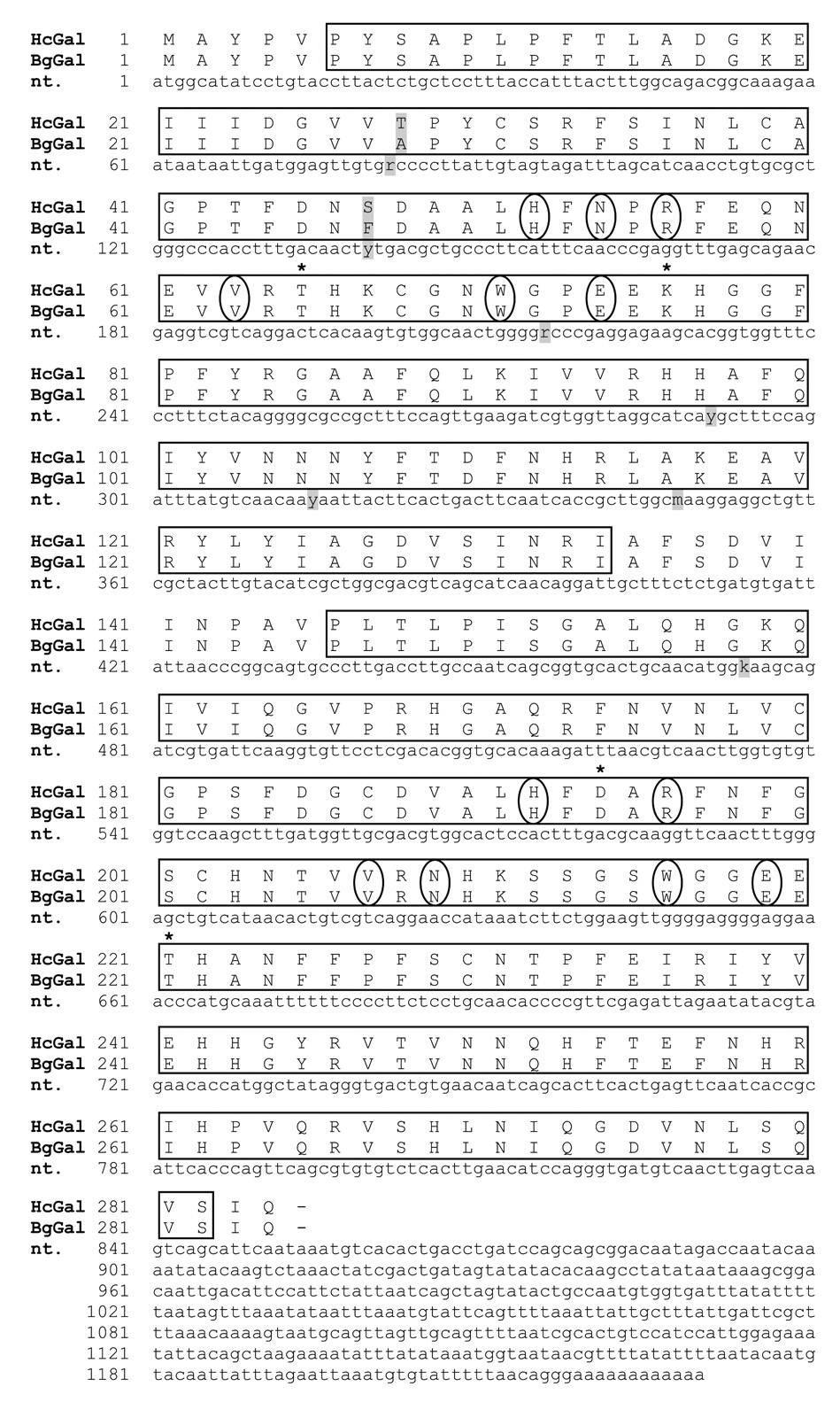
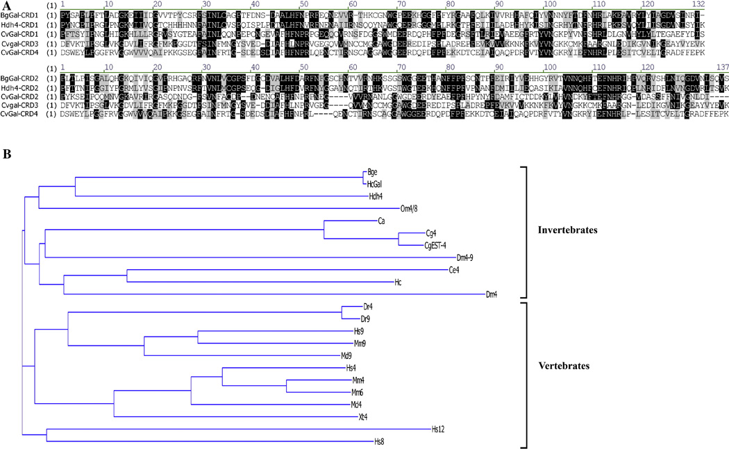
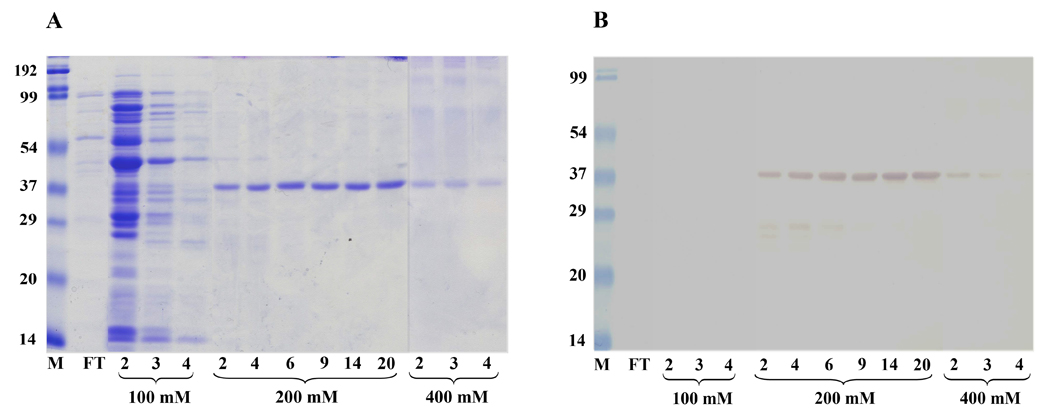
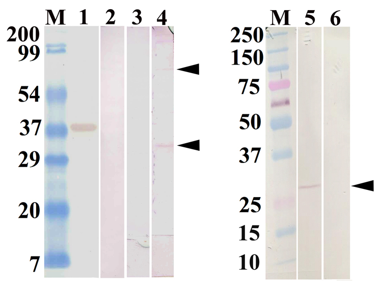
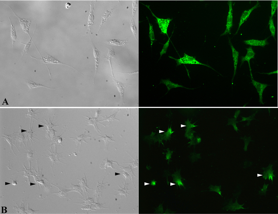
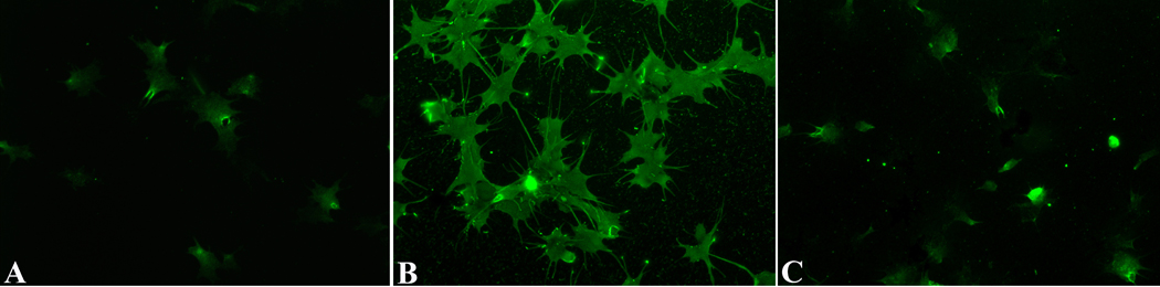
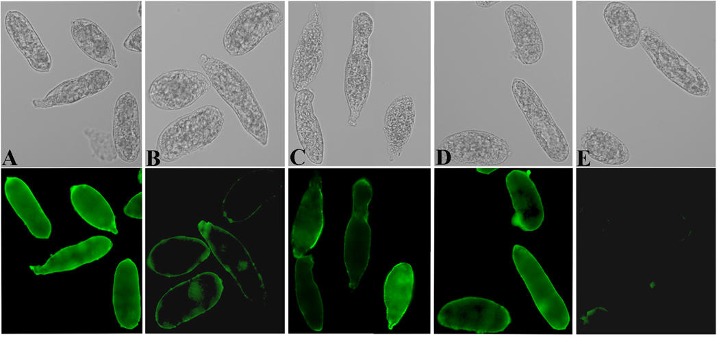
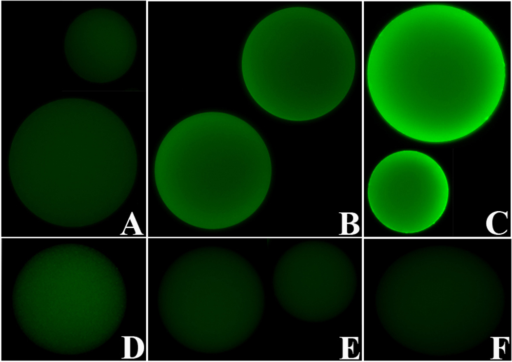
In the present study, a tandem-repeat type galectin was characterized from an embryonic cell line (Bge) and circulating hemocytes of the snail Biomphalaria glabrata, intermediate host of the human blood fluke Schistosoma mansoni. The predicted B. glabrata galectin (BgGal) protein of 32 kDa possessed 2 carbohydrate recognition domains, each displaying 6 of 8 conserved amino acids involved in galactoside-binding activity. A recombinant BgGal (rBgGal) demonstrated hemagglutinating activity against rabbit erythrocytes, which was specifically inhibited by galactose-containing sugars (lacNAc/lac>galNAc/gal). Although native galectin was immunolocalized in the cytoplasm of Bge cells and the plasma membrane of a subset of snail hemocytes (60%), it was not detected in cell-free plasma by Western blot analysis. The findings that rBgGal selectively recognizes the schistosome-related sugar, lacNAc, and strongly binds to hemocytes and the tegument of S. mansoni sporocysts in a sugar-inhibitable fashion suggest that hemocyte-bound galectin may be serving as a pattern recognition receptor for this, or other pathogens possessing appropriate sugar ligands. Based on molecular and functional features, BgGal represents an authentic galectin, the first to be fully characterized in the medically-important molluscan Class Gastropoda.


Figures

Fig. 1
Fig. 1
Structural characterization and comparison of the tandem-repeat galectins from the B. glabrata embryonic cell line (BgeGal) and circulating hemocytes (HcGal). Two CRDs were identified (see boxed areas for CRD1: aa6–aa134 and CRD2: aa146–aa282), separated by an 11 aa peptide linker. Comparison of nucleotide (nt) sequences show 5 differences (shaded in gray; r = a or g, y = c or t, m = a or c, k = g or t), that result in 2 aa changes (aa28 and aa47) within CRD1. Circled aa show 6 of 8 highly conserved aa within each CRD, responsible for the galactosyl-binding activity (CRD1: H52, N54, R56, V63, W71, E74; CRD2: H192, R196, V207, N209, W216, E219). Asterisks (*) indicate the 4 substituted aa (CRD1: T65, K76, CRD2: D194, T221).
Fig. 2
Fig. 2
(A) Protein sequence alignment of multi-CRD molluscan galectins: B. glabrata (BgGal), abalone Haliotis (Hdh4) and the oyster C. virginica (CvGal) (GenBank™ accession nos. ABQ09359, ABN54798 and DQ779197, respectively). First alignment series shows the aa identities (shaded in black) and similarities (gray) of the BgGal CRD1 with all other CRD1s, and CRD3 and 4 from the oyster. Second alignment series compares BgGal CRD2 with the other CRD2 sequences and the oyster CRD3 and 4. (B) Cluster tree analysis of selected invertebrate (Bge cells and B. glabrata hemocyte galectins, BgGal, HcGal, respectively; abalone Haliotis discus hannai Gal-4, Hdh4; Ornithodoros moubata Gal-4/8, Om4/8; D. melanogaster Gal-4/9, Dm4/9; C. elegans Gal-4, Ce4; Haemonchus contortus dual-CRD, Hc; Crassostrea spp., CgEST4, Cg4, Ca) and vertebrate (Danio rerio Gal-4 and 9, Dr4 and Dr9; Mus musculus Gal-4, 6 and 9, Mm4, Mm6 and Mm9; Monodelphis domestica Gal-4 and 9, Md4 and Md9; Xenopus tropicalis Gal-4, Xt4; Homo sapien Gal-4, 8, 9 and 12, Hs4, Hs8, Hs9 and Hs12) dual or tandem-repeat type galectins. This cluster analysis demonstrates the higher structural similarities within invertebrate tandem-repeat galectins compared to their vertebrate counterparts. GenBank™ accession nos.: Om4/8: BAF43802; Dm4: AAF57667; Dm4/9: AAL87743; Ca: ABC69709; Cg4: CAD79473; CgEST4: BG467428; Ce4: NP_497763; Hc: AAF63405; Dr4: AAR84192; Dr9: NP_001001817; Mm4: AAH94008; Mm6: O54891; Mm9: AAH03754; Md4: XM_001362670; Md9: XP_001375560; Xt4: NP_001011449; HS4: NP_006140; Hs8: AAF19370; HS9: AAI05945; HS12: NP_149092;
Fig. 3
Fig. 3
Comassie blue-stained SDS-PAGE gel displaying protein patterns of selected elution fractions taken during the purification of rBgGal on the His-trap column (A). After flow-through (FT), 100 mM imidazole elutions (lanes 2, 3 and 4) show release of many different sized proteins. Elution with 200 mM imidazole released a single 37 kDa protein corresponding to the estimated molecular mass of rBgGal (lanes 2, 4, 6, 9, 14, 20), while final elutions with 400 mM imidazole show a 37 kDa protein and other proteins. (B) Corresponding Western blot of proteins transferred from gel shown in Fig. 3A, probed with anti-His antibody and demonstrating reactivity with the 37 kDa protein eluted with 200 and 400 mM imidazole buffer, confirming that the 37 kDa eluted is the expressed-His-tagged rBgGal protein.
Fig. 4
Fig. 4
Western blots of SDS-PAGE-separated recombinant and immunoreactive endogenous BgGal probed with an anti-rBgGal polyclonal antiserum. 1: positive control containing the 37 kDa rBgGal only (arrow); 2: negative control containing blotted rBgGal probed with the anti-rBgGal antiserum previously absorbed with rBgGal; 3: Bge cell protein extract showing negative reactivity with the rabbit pre-immune serum; 4: reactivity of anti-rBgGal with 2 Bge cell derived proteins,at 32 kDa and 70 kDa (arrows); 5: immunoreactive of a 32 kDa protein (arrow) from a B. glabrata hemocyte homogenate; 6: B. glabrata cell-free hemolymph (plasma) sample showing negative reactivity with the rBgGal antiserum.
Fig. 5
Fig. 5
Differential interference contrast (DIC) and immunofluorescence images of Bge cells (A) and hemocytes (B) following treatment with the anti-rBgGal antibody and Alexa 488-conjugated secondary antibody. Bge cells show uniform distribution of the endogenous galectin within the cytoplasm, although the intensity of staining is variable. By contrast, only ~60% of circulating hemocytes expressed native galectin, with compartmentalization within the cytoplasm, and along the margins of filapodial membranes (arrows). (400X).
Fig. 6
Fig. 6
In vitro binding of rBgGal to the surface of Biomphalaria glabrata hemocytes. Live hemocytes, preincubated with rBgGal followed by fixation and treatement with anti-rBgGal antibody demonstrates intense fluorescent surface staining (B) compared to endogenous BgGal staining (A) or staining following exposure of hemocytes to rBgGal in the presence of 40 mM lactose (C). Lactose inhibition of the rBgGal binding to hemocytes demonstrates the presence of specific BgGal counterreceptors (ligands) on the surface of hemocytes.
Fig. 7
Fig. 7
DIC and immunofluorescence image of S. mansoni sporocysts pretreated with rBgGal followed by anti-rBgGal antibodies showing the binding of rBgGal to the sporocyst tegument (A). Partial inhibition of rBgGal binding by lactose (B) or lacNAc (C), but not fucose (D) demonstrates the specificity of rBgGal binding to sporocyst surface. Fig. 7E serves as a control, showing a complete absence of nonspecific or endogenous galectin-like proteins in sporocysts using the antirBgGal antibody (200X).
Fig. 8
Fig. 8
Immunofluorescence images of BSA-conjugated sepharose beads demonstrating the binding of rBgGal to lacNAc (LN) (B, E) and sialyated lacNAc (sLN) (C, F), but not to beads conjugated with BSA-LeX (D) or BSA alone (A).) Inhibition of the binding by lactose confirmed the specificity of rBgGal for LN and sLN (E, F). All immunofluorescent images were normalized to background control beads levels using Metamorph software, v. 7.2. (200X).

References

    1. Acosta-Rodriguez EV, Montes CL, Motran CC, Zuniga EI, Liu F-T, Rabinovich GA, Gruppi A. Galectin-3 mediates IL-4-induced survival and differentiation of B cells: functional cross-talk and implications during Trypanosoma cruzi infection. J. Immunol. 2004;172:493–502. - PubMed
    1. Adema CM, Arguello DF, II, Stricker SA, Loker ES. A family of fibrinogen-related proteins that precipitate parasite-derived molecules is produced by an invertebrate after infection. Proc. Natl. Acad. Sci. (USA) 1994;94:8691–8696. - PMC - PubMed
    1. Baldo BA, Uhlenbruck G. Anti-galactan activity in Tridacna maxima (Roding) haemolymph: Calcium dependence of the haemagglutinins and precipitins. Immunology. 1975;29:1161–1170. - PMC - PubMed
    1. Barat-Houari M, Hilliou F, Jousset F-X, Sofer L, Deleury E, Rocher J, Ravallec M, Galibert L, Delobel P, Feyereisen R, Fournier P, Volkoff A-N. Gene expression profiling of Spodoptera frugiperda hemocytes and fat body using cDNA microarray reveals polydnavirus-associated variations in lepidoperan host gene transcript levels. BMC Genomics. 2006;7:160–179. - PMC - PubMed
    1. Barondes SH, Cooper DNW, Gitt MA, Leffler H. Galectins: Structure and function of a large family of animal lectins. J. Biol. Chem. 1994;269:20807–20810. - PubMed

Publication types

MeSH terms