

Osteocytes, mechanosensing and Wnt signaling
- PMID: 18280232
- PMCID: PMC2349095
- DOI: 10.1016/j.bone.2007.12.224
Osteocytes, mechanosensing and Wnt signaling
Abstract
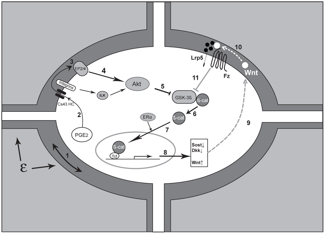
The majority of bone cell biology focuses on activity on the surface of the bone with little attention paid to the activity that occurs below the surface. However, with recent new discoveries, osteocytes, cells embedded within the mineralized matrix of bone, are becoming the target of intensive investigation. In this article, the distinctions between osteoblasts and their descendants, osteocytes, are reviewed. Osteoblasts are defined as cells that make bone matrix and osteocytes are thought to translate mechanical loading into biochemical signals that affect bone (re)modeling. Osteoblasts and osteocytes should have similarities as would be expected of cells of the same lineage, yet these cells also have distinct differences, particularly in their responses to mechanical loading and utilization of the various biochemical pathways to accomplish their respective functions. For example, the Wnt/beta-catenin signaling pathway is now recognized as an important regulator of bone mass and bone cell functions. This pathway is important in osteoblasts for differentiation, proliferation and the synthesis bone matrix, whereas osteocytes appear to use the Wnt/beta-catenin pathway to transmit signals of mechanical loading to cells on the bone surface. New emerging evidence suggests that the Wnt/beta-catenin pathway in osteocytes may be triggered by crosstalk with the prostaglandin pathway in response to loading which then leads to a decrease in expression of negative regulators of the pathway such as Sost and Dkk1. The study of osteocyte biology is becoming an intense area of research interest and this review will examine some of the recent findings that are reshaping our understanding of bone/bone cell biology.
Figures
References
-
- Ahmed Y, Hayashi S, Levine A, Wieschaus E. Regulation of Armadillo by a Drosophila APC Inhibits Neuronal Apoptosis during Retinal Development. Cell. 1998;93:1171–1182. - PubMed
-
- Almeida M, Han L, Lowe V, Warren A, Kousteni S, O'Brien CA, Manolagas S. Reactive Oxygen Species Antagonize the Skeletal Effects of Wnt/β-Catenin In Vitro and Aging Mice by Diverting β-Catenin from TCF- to FOXO-Mediated Transcription. J Bone Min Res. 2006;21 Suppl 1:S26. (abst 1092)
-
- Anderson EJ, Kaliyamoorthy S, Iwan J, Alexander D, Knothe Tate ML. Nano-microscale models of periosteocytic flow show differences in stresses imparted to cell body and processes. Ann Biomed Eng. 2005;33:52–62. - PubMed
Publication types
MeSH terms
Substances
Grants and funding
LinkOut - more resources
Full Text Sources
Other Literature Sources
Medical
