. 2008 Feb 15;29(3):291-301.
doi: 10.1016/j.molcel.2007.10.036.

Beta clamp directs localization of mismatch repair in Bacillus subtilis

Affiliations

Beta clamp directs localization of mismatch repair in Bacillus subtilis

Lyle A Simmons et al. Mol Cell. .

Abstract

MutS homologs function in several cellular pathways including mismatch repair (MMR), the process by which mismatches introduced during DNA replication are corrected. We demonstrate that the C terminus of Bacillus subtilis MutS is necessary for an interaction with beta clamp. This interaction is required for MutS-GFP focus formation in response to mismatches. Reciprocally, we show that a mutant of the beta clamp causes elevated mutation frequencies and is reduced for MutS-GFP focus formation. MutS mutants defective for interaction with beta clamp failed to support the next step of MMR, MutL-GFP focus formation. We conclude that the interaction between MutS and beta is the major molecular interaction facilitating focus formation and that beta clamp aids in the stabilization of MutS at a mismatch in vivo. The striking ability of the MutS C terminus to direct focus formation at replisomes by itself, suggests that it is mismatch recognition that licenses MutS's interaction with beta clamp.


Figures

Figure 1
Figure 1. MutS interacts with the β-clamp through the C-terminal 58 amino acids
The peptide array and far western blot were probed with β-clamp bearing a single Myc tag (β-Myc). (A) Shown are 10mer peptides of interest from the MutS N-terminus that failed to bind β or the C-terminal 58 amino acids that bound β-Myc. The amino acid sequence is indicated and the β-clamp binding motif or N-terminal motif is highlighted in grey. Ponceau staining of the peptides is shown in the left most panel. (B) β-Myc, MutS, MutS800 and His-DnaE probed with β-Myc. (C) Interaction between MutS or MutS800 with β-clamp covalently linked to a sensor chip was completed using surface plasmon resonance as measured with a BIAcore biosensor (“Experimental Procedures”). Representative SPR traces for MutS 1.0 μM (blue) and 4.0 μM (red); MutS800 2.0 μM (green), 10.0 μM (purple) are shown.
Figure 2
Figure 2. The MutS C-terminus is required for MMR activity in vivo
(A) Diagram of B. subtilis MutS domain structure and segments of the protein absent from the truncated mutants. The domains were determined by alignment with Thermus aquaticus MutS and the domain nomenclature follows that of the crystal structure (Obmolova et al., 2000). Briefly, domains I and IV are involved in mismatch binding, domain III is the central core of the protein, domain II resembles an RNase H-like fold and domain V is an ABC ATPase. (B) Corresponding mutation frequencies associated with strains lacking the indicated C-terminal amino acids of MutS. Relative stability of the resultant proteins was determined by immunoblot analysis and are presented above the histogram. (C) Mutation frequency resulting from alanine scanning mutagenesis of the MutS β-clamp binding motif. An immunoblot of the in vivo stability of the resulting proteins is also shown. The alanines substitutions in the MutS β-clamp binding motif are indicated in bold type. Error bars represent the standard deviation from at least triplicate experiments.
Figure 3
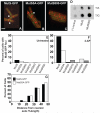
Figure 3. The C-terminus of MutS is required for focus formation
All samples shown are representative. Cells were treated with 600 μg/mL 2-AP to induce mismatches. Shown are (A) MutS-GFP foci; (B) MutS5A-GFP; (C) MutS800-GFP. (D) MutS, MutS800 and (-) control (no protein) were incubated with homoduplex DNA (T/A) or mismatched heteroduplex DNA (T/G) as described (Biswas and Hsieh, 1996). MutS bound to DNA was retained on the nitrocellulose membrane and exposed to film. (E) Percent of cells with foci untreated. (F) Percent of cells with foci 45 min following addition of 2-AP. (G) Comparison of the cellular position of MutS-GFP single foci (black bar) and MutS5A-GFP foci (white bar) following 2-AP addition. The number of cells scored for untreated and 2-AP treated in panels D and E are as follows: MutS-GFP (n=837, n=1309); MutS5A-GFP (n=1044, n=1102); MutS800-GFP (n=396, n=958); MutS810-GFP (n=567, n=410); MutS830-GFP (n= 382, n= 944); MutS840-GFP (n=595, n=1022), MutS850-GFP (n=771, n=801). The cell membrane is stained with FM4-64 and is colored in red, while MutS-GFP foci are in green. The white bar indicates 2μm. White dots are adjacent to MutS-GFP foci.
Figure 4
Figure 4. MutS mutant proteins are impaired for supporting MutL-GFP focus formation
Cells were grown in S750 minimal medium and treated with 600 μg/mL 2-AP to induce mismatches 45 min prior to visualization. Cell images shown are representative of several experiments. Shown are (A) MutL-GFP in a wild type mutS background; (B) MutL-GFP in a mutS5A (β-clamp binding motif replaced with alanines) background; (C) MutL-GFP in a mutS800 background; (D) MutL-GFP in a ΔmutS background. (E) Percent of cells with MutL-GFP foci untreated. (F) Percent of cells with MutL-GFP foci 45 min following the addition of 2-AP. (G) Comparison of the subcellular position of MutL-GFP foci in a mutS (black bars) and mutS5A (white bars) genetic background (n=150). Number of cells scored for the graphs in panels E and F with the untreated number followed by the 2-AP treated number are as follows: MutL-GFP (n=1396, n=891) in a wild type mutS background; (n=1028, n=1042) in a ΔmutS background; (n=2543, n=2866) in a mutS800 background; (n=1142, n=884) in a mutS5A background.
Figure 5
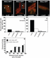
Figure 5. The C-terminus of MutS is both necessary and sufficient for focus formation
YFP was expressed from the amyE locus and visualized by fluorescence microscopy. Shown are (A) YFP and membrane; (B) MutSc-YFP in a mutSL- strain and membrane; (C) MutSC-YFP and membrane in a wild type strain; (D) MutSC-YFP and membrane in a mutL- strain. The white bar indicates 2μm. The percent of cells with foci 45 min after addition of IPTG are shown in each panel and the numbers of cells scored are indicated in each panel. (E) Representative β-clamp-GFP foci are shown during normal growth in S750 minimal medium. (F) Subcellular position of MutSC-YFP (white bars), β-clamp-GFP (grey bars) and DnaX-GFP (black bars) are presented as the percent of cells versus the distance from the nearest pole.
Figure 6
Figure 6. A mutant form of the β-clamp is reduced for supporting MutS-GFP formation
(A) Relative mutation frequency of the βG73R strain compared with MMR deficient strains at 30°C, and (B) 37°C. (C) Homology model of B. subtilis β-clamp with the G73R substitution shown (green). Residues colored in red and yellow are conserved with residues in E. coli β that comprise the hydrophobic pocket (red) and conserved residues important for interaction between E. coli clamp loader component δ’ and β (yellow). (D) Enlargement of the region containing the G73R mutation (green). (E) Immunoblot of β and βG73R protein levels in whole cell lysates at 30°C and 37°C. Protein load was normalized to cell number. β is in lanes 1 and 3 and βG73R is in lanes 2 and 4. (F) Quantitation of MutS-GFP focus formation in wild type and βG73R backgrounds at 30°C and 37°C.

Similar articles

Cited by

References

    1. Acharya S, Foster PL, Brooks P, Fishel R. The coordinated functions of the E. coli MutS and MutL proteins in mismatch repair. Mol Cell. 2003;12:233–246. - PubMed
    1. Acharya S, Wilson T, Gradia S, Kane MF, Guerrette S, Marsischky GT, Kolodner R, Fishel R. hMSH2 forms specific mispair-binding complexes with hMSH3 and hMSH6. Proc Natl Acad Sci U S A. 1996;93:13629–13634. - PMC - PubMed
    1. Allen DJ, Makhov A, Grilley M, Taylor J, Thresher R, Modrich P, Griffith JD. MutS mediates heteroduplex loop formation by a translocation mechanism. Embo J. 1997;16:4467–4476. - PMC - PubMed
    1. Beuning PJ, Sawicka D, Barsky D, Walker GC. Two processivity clamp interactions differentially alter the dual activities of UmuC. Mol Microbiol. 2006;59:460–474. - PubMed
    1. Biswas I, Hsieh P. Identification and characterization of a thermostable MutS homolog from Thermus aquaticus. J Biol Chem. 1996;271:5040–5048. - PubMed

Publication types

MeSH terms

Substances