. 2008 Feb 15;29(3):362-75.
doi: 10.1016/j.molcel.2007.12.024.

Ran-binding protein 3 phosphorylation links the Ras and PI3-kinase pathways to nucleocytoplasmic transport

Affiliations

Ran-binding protein 3 phosphorylation links the Ras and PI3-kinase pathways to nucleocytoplasmic transport

Sang-Oh Yoon et al. Mol Cell. .

Abstract

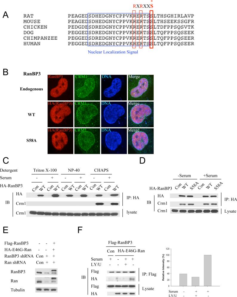
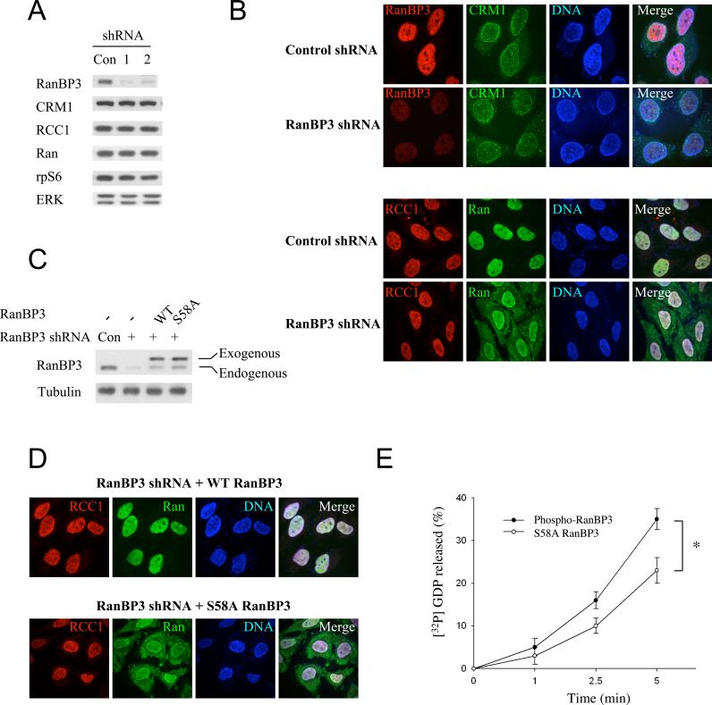
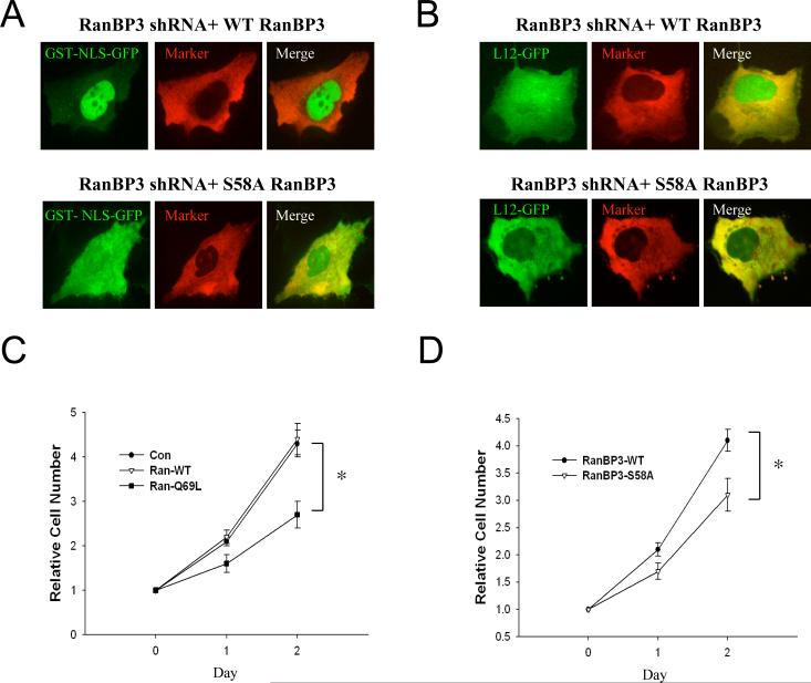
The major participants of the Ras/ERK and PI3-kinase (PI3K) pathways are well characterized. The cellular response to activation of these pathways, however, can vary dramatically. How differences in signal strength, timing, spatial location, and cellular context promote specific cell-fate decisions remains unclear. Nuclear transport processes can have a major impact on the determination of cell fate; however, little is known regarding how nuclear transport is regulated by or regulates these pathways. Here we show that RSK and Akt, which are activated downstream of Ras/ERK and PI3K, respectively, modulate the Ran gradient and nuclear transport by interacting with, phosphorylating, and regulating Ran-binding protein 3 (RanBP3) function. Our findings highlight an important link between two major cell-fate determinants: nuclear transport and the Ras/ERK/RSK and PI3K/Akt signaling pathways.


Figures

Fig. 1
Fig. 1
RanBP3 is a RSK binding partner. (A) Schematic representation of RSK structure (B) The partial RanBP3 clones (8, 32, and 41) obtained from the two-hybrid mouse library screen are schematically aligned with the two full-length human clones (isoforms RanBP3a and RanBP3b). (C) Clone 8 obtained from two-hybrid screening was transformed into yeast with plasmids bearing the indicated GAL4 DNA binding domain fusions and grown on minimal media lacking uracil, leucine, and adenine to select for two-hybrid interaction. Clone SKAR, which is known to interact with p70S6K1, was used as a positive control. (D) HEK293 cells were transfected with either HA-RSK or control vector with FLAG-RanBP3 for 24 h, and then incubated in serum-free media for an additional 20 h. Cells were treated with 50 ng/ml PMA for 15 min and lysed with either CHAPS or Triton X-100 lysis buffer in the presence or absence of DSP, a cross-linking reagent. Immunoprecipitation and immunoblot analysis were performed. The arrowhead indicates the HA-RSK co-immunoprecipitating FLAG-tagged RanBP3.
Fig. 2
Fig. 2
RSK phosphorylates RanBP3 at Serine 58 residue in vitro and in vivo. (A) Schematic representation of RanBP3. RanBP3 has a Nuclear Localization Signal (NLS), a Ran Binding Domain (RBD), and possible RSK phosphorylation sites. (B) Low-energy collision-induced dissociation spectrum of the RanBP3 pS58 phosphopeptide. (C) HEK293 cells were transfected with either HA-WT-RSK1 or HA-KD-RSK1 (Δ/Δ RSK1) for 24 h, and then incubated in serum-free media for an additional 20 h. Cells were left untreated or treated with 50 ng/ml PMA for 15 min, and immunoprecipitation was performed. RSK phosphotransferase assays were performed with purified GST-WT RanBP3, GST-S57A RanBP3, and GST-S58A RanBP3. (D) HEK293 cells were transfected with HA-WT-RSK1 for 24 h, and then incubated in serum-free media for an additional 20 h. During the last 1 h, cells were left untreated or treated with 10 μM U0126. After treatment with 50 ng/ml PMA, immunoprecipitation and RSK phosphotransferase assays were performed. (E) HEK293 cells were transfected with control plasmid, WT RanBP3, S58A RanBP3 or S58D RanBP3 for 24 h, and incubated in serum-free media for an additional 20 h. After treatment with 50 ng/ml PMA for 15 min, cells were lysed and immunoblot analysis was performed. (F) HA-RanBP3 plasmid was co-transfected with either HA-RSK1 or HA-RSK2 into HEK293 cells for 24h, and then cells were incubated in serum-free medium for an additional 20 h. Cells were lysed and immunoblot analysis was performed. (G) RSK knockdown HEK293 cells were generated using RSK1 and RSK2 shRNA. Cells were incubated in serum-free media for 20 h. After treatment with 50 ng/ml PMA for 15 min, cells were lysed and immunoblot analysis was performed.
Fig. 3
Fig. 3
Akt phosphorylates RanBP3 at Serine 58 residue in vitro and in vivo. (A) HEK293 cells were transfected with either HA-WT Akt or HA-KD Akt for 24 h, and then incubated in serum-free media for an additional 20 h. Cells were left untreated or treated with 100 nM insulin for 15 min, and immunoprecipitation was performed. Akt phosphotransferase assays were performed using purified GST-WT RanBP3, GST-S57A RanBP3, GST-S58A RanBP3, and GST-S153A RanBP3 as substrate. (B) HEK293 cells were transfected with HA-WT Akt for 24 h, and then incubated in serum-free medium for an additional 20 h. During the last 1 h, cells were left untreated or treated with 20 μM LY294002. After treatment with 100 nM insulin, immunoprecipitation and Akt phosphotransferase assays were performed using GST-WT RanBP3, GST-S58A RanBP3, and GST-S153A RanBP3 as substrates. (C) HEK293 cells were transfected with either HA-Akt or control vector with FLAG-RanBP3 for 24 h, and then incubated in serum-free media for an additional 20 h. Cells were treated with 100 nM insulin for 15 min, and lysed with either CHAPS- or Triton X-100 –containing buffer in the presence or absence of DSP, a cross-linking reagent. Immunoprecipitation and immunoblot analysis were perfomred. The arrowheads indicate the HA-Akt co-immunoprecipitating FLAG-tagged RanBP3. (D) HEK293 cells were transfected with HA-Akt for 24 h, and then incubated in serum-free medium for an additional 20 h. Cells were lysed and immunoblot analysis was performed. (E) Akt knockdown HEK293 cells were generated using Akt1 and Akt2 shRNA. Cells were incubated in serum-free media for 20 h. After treatment with 100 nM insulin for 15 min, cells were lysed and immunoblot analysis was performed. (F) HEK293 cells were incubated in serum-free media for 20 h. During the last 1 h, cells were treated with pleckstrin homology domain-dependent Akt inhibitor. After treatment with 100 nM insulin for 15 min, cells were lysed and immunoblot analysis was performed.
Fig. 4
Fig. 4
Growth factors regulate RanBP3 phosphorylation in PI3K/Akt- and ERK/RSK-dependent pathways. (A-F) HEK293 cells were incubated in serum-free media for 20 h. During the last 1 h, cells were treated with inhibitors (10 μM U0126, 20 μM LY294002, or 20 ng/ml rapamycin). After treatment with indicated agonists (50 ng/ml PMA, 100 nM insulin, or 50 ng/ml EGF) for 15 min, cells were lysed and immunoblot analysis was performed.
Fig. 5
Fig. 5
RanBP3 phosphorylation does not affect RanBP3 localization and binding affinity toward Crm1. (A) Alignment of RanBP3 protein sequences in various mammals. (B) For the detection of endogenous RanBP3 localization in cells, HeLa cells were stained with anti-RanBP3 antibody, and subjected to confocal microscopy. For exogenous RanBP3 detection, HeLa cells were transfected with either HA-WT RanBP3 or HA-S58A RanBP3, and stained with anti-HA antibody. The localization of these proteins was visualized by confocal microscopy. (C) HEK293 cells were transfected with either HA-RanBP3 or control plasmid for 24 h, and then incubated in serum-free media for an additional 20 h. Cells were left untreated or treated with 20% serum for 15 min, and lysed with Triton X-100, NP-40, or CHAPS buffer. Immunoprecipitation and immunblot analysis were performed. (D) HEK293 cells were transfected with control plasmid, HA-WT RanBP3, or HA-S58A RanBP3 for 24 h, and then incubated in serum-free media for an additional 20 h. Cells were left untreated or treated with 20% serum, and immunoprecipitation and immunoblot analysis were performed. (E) RanBP3 and Ran double-knockdown HEK293 cells were generated using RanBP3 and Ran shRNA. Knockdown cells were transfected with RanBP3 and E46G-Ran, and immunoblot analysis was performed. (F) RanBP3 and Ran double-knockdown cells were transfected with either HA-E46G Ran or control vector plus Flga-RanBP3 for 24 h, and then incubated in serum-free media for an additional 20 h. During the last 1 h, cells were treated with inhibitors (10 μM U0126 and 20 μM LY294002). After incubation with 20% serum for 15 min, cells were lysed and immunoprecipitation was performed.
Fig. 6
Fig. 6
RanBP3 modulates the Ran gradient. (A) RanBP3 knockdown HeLa cells were generated as described in methods. Stable cells were lysed and immunoblot analysis was performed. (B) RanBP3 knockdown and control HeLa cells were stained with antibodies against RanBP3, CRM1, RCC1, and Ran. Localization of these proteins was visualized by confocal microscopy. (C) Stable WT RanBP3 and S58A RanBP3 HeLa cells were generated from RanBP3 knockdown cells and immunoblot analysis was performed. (D) Stable WT RanBP3 and S58A RanBP3 HeLa cells were stained with anti-RCC1 and anti-Ran antibodies, and subjected to confocal microscopy. (E) RanGEF (RCC1) assay was performed as described in methods. Data represent the mean±S.D. of three independent experiments. Results were statistically significant (*, p < 0.01) using Student's t test.
Fig. 7
Fig. 7
RanBP3 phosphorylation regulates nuclear transport. (A) GST-NLS-GFP and injection marker were injected into the cytoplasm of HeLa cells. After 10 min, cells were fixed and the injected proteins were visualized by confocal microscopy. (B) GFP-L12 and injection marker were injected into the cytoplasm of HeLa cells. After 3 min, cells were fixed and the injected proteins were visualized by confocal microscopy. (C) Cells were transfected with control vector or Ran constructs, and cell numbers were counted at the indicated times. Data represent the mean±S.D. of three independent experiments. Results were statistically significant (*, p < 0.01) using Student's t test. (D) RanBP3 knockdown cells expressing stable WT RanBP3 and S58A RanBP3 were plated, and cell numbers were counted at indicated times. Data represent the mean±S.D. of three independent experiments. Results were statistically significant (*, p < 0.01) using Student's t test.

References

    1. Anjum R, Roux PP, Ballif BA, Gygi SP, Blenis J. The tumor suppressor DAP kinase is a target of RSK-mediated survival signaling. Curr Biol. 2005;15:1762–1767. - PubMed
    1. Arnaoutov A, Azuma Y, Ribbeck K, Joseph J, Boyarchuk Y, Karpova T, McNally J, Dasso M. Crm1 is a mitotic effector of Ran-GTP in somatic cells. Nat Cell Biol. 2005;7:626–632. - PubMed
    1. Bonni A, Brunet A, West AE, Datta SR, Takasu MA, Greenberg ME. Cell survival promoted by the Ras-MAPK signaling pathway by transcription-dependent and -independent mechanisms. Science. 1999;286:1358–1362. - PubMed
    1. Cartlidge RA, Knebel A, Peggie M, Alexandrov A, Phizicky EM, Cohen P. The tRNA methylase METTL1 is phosphorylated and inactivated by PKB and RSK in vitro and in cells. Embo J. 2005;24:1696–1705. - PMC - PubMed
    1. Clarke PR. The Crm de la creme of mitosis. Nat Cell Biol. 2005;7:551–552. - PubMed

Publication types

MeSH terms

Substances