. 2008 Apr 1;316(1):6-20.
doi: 10.1016/j.ydbio.2007.12.040. Epub 2008 Jan 5.

Regulation of the development of tectal neurons and their projections by transcription factors Brn3a and Pax7

Affiliations

Regulation of the development of tectal neurons and their projections by transcription factors Brn3a and Pax7

Natalia Fedtsova et al. Dev Biol. .

Abstract

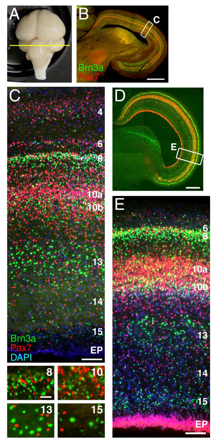
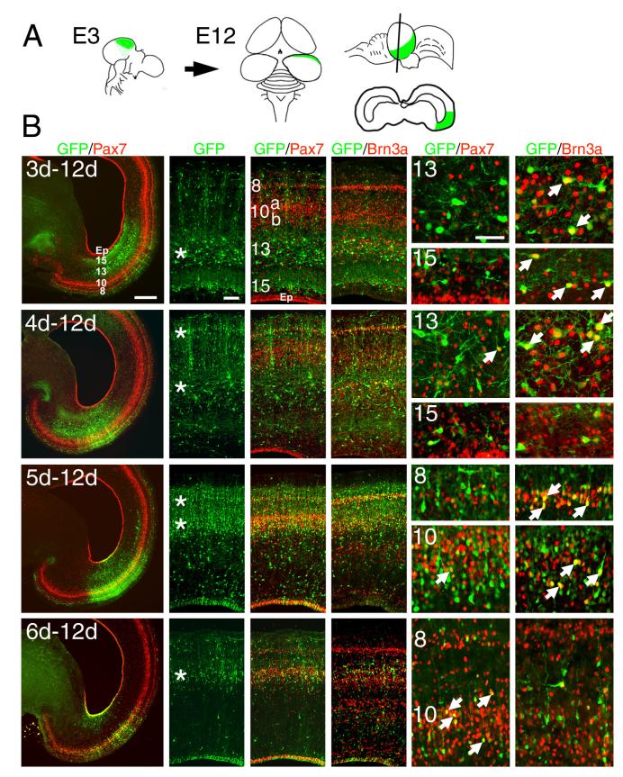
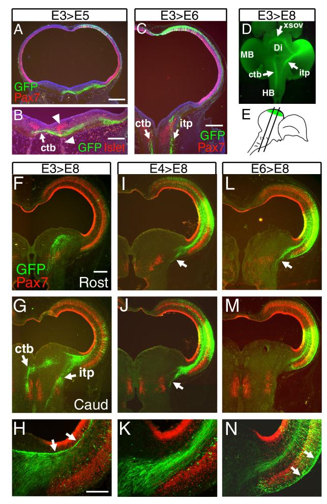
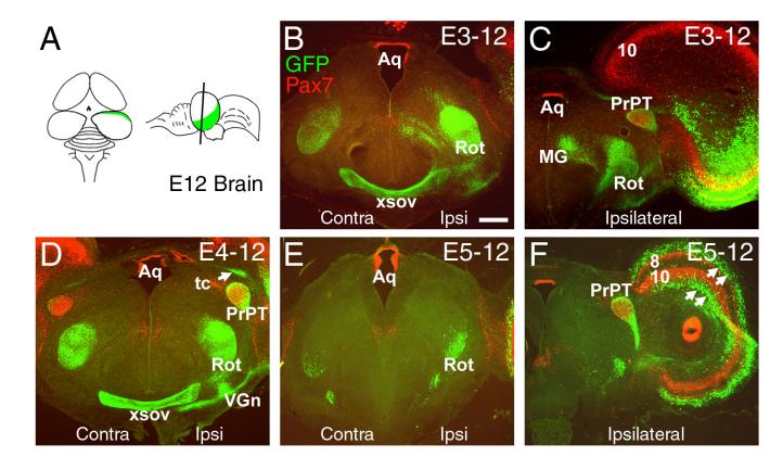
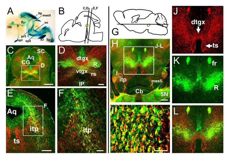
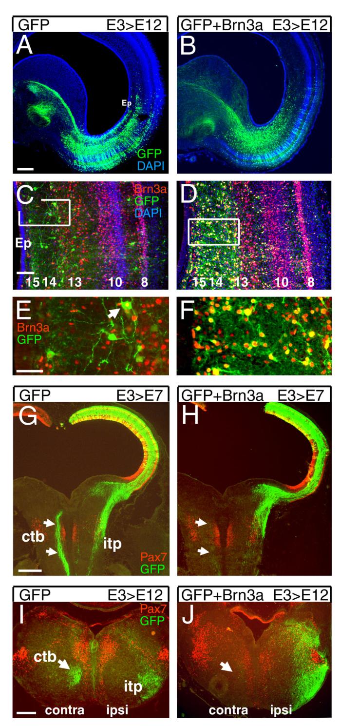
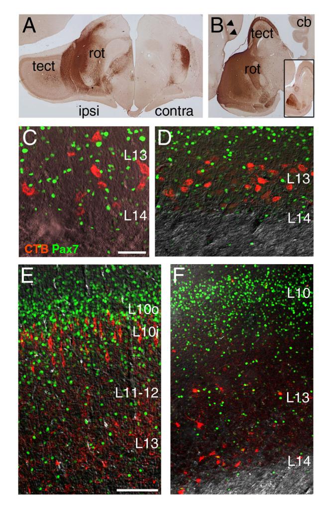
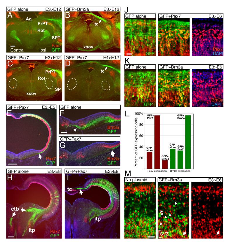
The rostral part of the dorsal midbrain, known as the superior colliculus in mammals or the optic tectum in birds, receives a substantial retinal input and plays a diverse and important role in sensorimotor integration. However, little is known about the development of specific subtypes of neurons in the tectum, particularly those which contribute tectofugal projections to the thalamus, isthmic region, and hindbrain. Here we show that two homeodomain transcription factors, Brn3a and Pax7, are expressed in mutually exclusive patterns in the developing and mature avian midbrain. Neurons expressing these factors are generated at characteristic developmental times, and have specific laminar fates within the tectum. In mice expressing betagalactosidase targeted to the Pou4f1 (Brn3a) locus, Brn3a-expressing neurons contribute to the ipsilateral but not the contralateral tectofugal projections to the hindbrain. Using misexpression of Brn3a and Pax7 by electroporation in the chick tectum, combined with GFP reporters, we show that Brn3a determines the laminar fate of subsets of tectal neurons. Furthermore, Brn3a regulates the development of neurons contributing to specific ascending and descending tectofugal pathways, while Pax7 globally represses the development of tectofugal projections to nearly all brain structures.


Figures

Figure 1
Figure 1. Lamina-specific expression of Brn3a and Pax7 in the chick tectum
The expression patterns of Brn3a and Pax7 was examined in a 5 week-old chick (A-C) and in an E12 chick embryo (D-E). (A) An intact 5 week chick brain showing the plane of section in subsequent views. (B) Low magnification view of the tectum with immunofluorescent staining for Brn3a and Pax7. (C) Laminar distribution of Brn3a and Pax7 neurons in the mature tectum (rotated relative to B, as shown by inset box). High magnification views of layers 8, 10, 13 and 15 demonstrate that Brn3a and Pax7 are not co-expressed at the cellular level. (D,E) Expression of Brn3a and Pax7 at E12, at which time the laminar expression pattern of these markers has been established, and all of the principal deep layers of the tectum can be identified. In C and E counterstaining with DAPI is used to identify all nuclei. Tectal lamina are designated numerically; EP, ependymal layer. View E is rotated relative to D, as shown by inset box. The nomenclature for the tectal layers used here, derived from the work of Ramon y Cajal (1995), is directly related to an alternate system in which layer 1 corresponds to the stratum opticum (SO), layers 2-11 to the stratum griseum et fibrosum superficiale (SGFS), sublayers a-j, layer 13 to the stratum griseum centrale (SGC), layer 14 to the stratum album centrale (SAC) and layer 15 to the stratum griseum et fibrosum periventriculare (SGFP, LaVail and Cowan, 1971a). Scale: B, 1mm; C, 100μm (insets, 25μm); D, 500μm; E, 50μm.
Figure 2
Figure 2. Birthdating of Brn3a and Pax7 expressing tectal neurons
A plasmid encoding a GFP reporter was electroporated unilaterally into the tectal neuroepithelium at each day from E3 to E6, and the electroporated tecta were analyzed at E12 for GFP, Brn3a and Pax7 expression. In each case, strong expression of the GFP marker indicates neurons which exited the cell cycle (i.e. were “born”) shortly after electroporation. (A) Overall strategy for the electroporation experiments. Rotation of the tectum between E6 and E12 results in a shift of the dorsal/rostral electroporated area at E3-E5 to a lateral position by E12 (Thanos and Mey, 2001). However, the region of electroporation can be recognized at any stage by the GFP-labeled radial processes of a subset of the electroporated cells, and a fraction of labeled cells that remain in the ependymal zone. (B) Brn3a and Pax7 expressing neurons generated at E3-E6, and analyzed at E12. In the views showing GFP expression alone, asterisks indicate the layers with the greatest numbers of neurons born soon after the electroporation. In the high magnification views, arrows indicate cells co-expressing GFP and the indicated transcription factor. The predominant pattern of neurogenesis is for newly born cells to be generated first in the in deep layers (L13-L15) at E3-E4, then in superficial layers (L8) at E4-E5, and finally in the intermediate layer (L10) at E5-E6. In layer 10, the peak generation of Brn3a-expressing cells (E5) appears to precede that of the Pax7-expressing population (E6). Data shown are representative of at least 5 cases electroporated at each developmental stage. Scale: B, low power, 500μm; medium power, 100μm; high power, 50μm.
Figure 3
Figure 3. Genesis of the descending tectofugal tracts
Neurons contributing to the tectofugal tracts were marked by the electroporation of a plasmid encoding a GFP reporter into the tectal neuroepithelium at various stages from E3 to E6. (A,B) Embryos electroporated at E3 and examined at E5. Axons of the crossed tectobulbar (CTB) pathway are strongly labeled, and have crossed the midline by E5. (B) Co-labeling with GFP and Islet1/2 shows the relationship of the decussation of the CTB (arrow) to the developing oculomotor neurons and tract (arrowheads). (C) Embryos electroporated at E3 and examined at E6. Descending fibers of the CTB and ipsilateral tectopontine tract (ITP) can be easily discerned by this stage. (D) Ventral, whole-mount view of GFP fluorescence in a brain electroporated at E3 and examined at E8, showing the electroporated region of the tectum and projections to hindbrain and diencephalon. (E) Diagram depicting the region of electroporation and plane of section for subsequent views of the midbrain and hindbrain. (F-N) Descending tectofugal axons from embryos electroporated at stages from E3-E6, and examined at E8. (F-H) In embryos electroporated at E3, labeled axons are prominent in both the CTB and ITP. Some of the early-generated neurons contribute to axons which fasciculate in the deepest tectal lamina, adjacent to the ependymal layer (arrows, H). (I-K) In embryos electroporated at E4 axons of the ITP continue to be strongly labeled (arrows I,J), but the generation of neurons contributing to the CTB is nearly complete. Similar results were obtained at E5 (not shown). (L-N) By E6 the generation of neurons contributing to the descending pathways is nearly complete. Late born neurons migrate extensively and send axons tangentially, superficial to layer 10 (arrows, N). Views A-C are counterstained with DAPI to reveal all nuclei. In F-H, the cell bodies of most of the electroporated neurons are out of the plane of section. In low power views of the tectum at E8 (F,G,I,J,L,M), GFP expression does not appear lamina-specific as it does in Figure 2 (E12) because the migration of neurons to specific layers is ongoing at E8, and because the long exposure times needed to reveal distant axon projections obscure the lamina-specific expression of GFP within the tectum. 10, future tectal layer 10; Ctb, crossed tectobulbar tract; Di, diencephalon; Ep, ependymal layer; HB, hindbrain; itp, ipsilateral tectopontine tract; MB, midbrain; xsov, ventral supraoptic decussation. Data shown are representative of at least 5 cases at E3 and E4 and 3 cases at E6. Scale: A, 400μm; B, 200μm; C, 400μm; F, 500μm; H, 200μm.
Figure 4
Figure 4. Genesis of the ascending tectofugal tracts
A plasmid expressing a GFP reporter was electroporated into the tectal neuroepithelium at various stages from E3 to E5, and the ascending tectofugal tracts were examined at E12. (A) Plane of section for subsequent views. (B, C) Electroporation at E3 results in heavy labeling of the ipsilateral nucleus rotundus, and moderate labeling of the contralateral nucleus rotundus and the pretectal nucleus. Labeling of the medial geniculate nucleus (nucleus ovoidalis) in embryos electroporated at E3 varied between cases and probably results from incidental labeling of the inferior colliculus. Section (C) is caudal to (B). (D) Electroporation at E4 heavily labels the ipsilateral and contralateral nucleus rotundus, the pretectal nucleus, the ventral geniculate nucleus, the xsov and the tectal commissure. (E,F) Electroporation at E5 shows that generation of neurons contributing to the nucleus rotundus is nearly complete at this stage; some fibers innervating the area surrounding the principal pretectal nucleus continue to be heavily labeled. Section (F) is caudal to (E). Arrows in (F) indicate tectal neurons which have migrated laterally away from the area of electroporation in distinct zones superficial to layer 8 and deep to layer 10. Aq, aqueduct; MG, medial geniculate nucleus (nucleus ovoidalis); PrPT, principal pretectal nucleus; Rot, nucleus rotundus; 8, 10, tectal lamina; tc, tectal commissure; VGn, ventral geniculate nucleus (pregeniculate nucleus); xsov, ventral supraoptic decussation. Data shown represent at least 3 cases at each developmental stage. Scale 400μm.
Figure 5
Figure 5. Tectofugal projections of Brn3a neurons in the mouse
Mice expressing a LacZ marker targeted to the Brn3a locus were used to examine the tectofugal pathways. In all immunofluorescence views, βgalactosidase expressed from the Brn3a/tauLacZ allele is labeled in green, and intermediate neurofilament immunoreactivity appears in red. In accordance with accepted terminology in rodents (Paxinos and Watson, 2005) the crossed descending tract from the tectum is here referred to as the tectospinal tract (TS) rather than the CTB. (A) An E16.5 Brn3atauLacZ/+ embryo, hemisected at the midline, and stained for βgalactosidase activity with X-gal. The major tracts formed by Brn3a-expressing neurons, including the ITP and tract/neurons of the mesV, are shown. Brn3a neurons also contribute to the retinothalamic/retinocollicular pathway, the habenulopeduncular tract, the olivocerebellar tract, and trigeminal afferent fibers. (B) Sagital view of an E16.5 brain, showing the planes of section in views C-F. Location of the red nucleus is shown in yellow. (C,D) Coronal section of the E16.5 tectum at the level of the red nucleus. βgalactosidase immunoreactive fibers contribute to the rubrospinal tract but not the decussating fibers of the CTB. The inset box in C appears enlarged in D. (E,F) More rostral view of the tectofugal pathways showing βgalactosidase expression in the ITP, but not the TS. The inset box in E appears enlarged in F. (G) Schematic of the postnatal (P10) brain showing plane of section for subsequent views. (H-L) Horizontal section of the postnatal brain of a Brn3atauLacZ/+ mouse at the level of the dorsal tegmental decussation shows expression of βgalactosidase in the red nucleus and ITP, but not the decussating fibers of the TS. The small inset box in H appears enlarged in I to show co-localization of neurofilament and LacZ staining in the ITP; the large inset box in H appears enlarged in J-L. 5N, trigeminal nucleus; Aq, aqueduct; Cb, cerebellum; CG, central gray; dtgx, dorsal tegmental decussation; fr, fasciculus retroflexus; Hb, habenula; IO, inferior olive; IP, interpeduncular nucleus; itp, ipsilateral tectopontine tract; mes5, mesencephalic trigeminal (and associated tract); oc, olivocerebellar tract; opt, optic tract; pr5, principal tract of trigeminal; R, red nucleus; rs, rubrospinal tract; Sp, spinal cord; sp5, spinal tract of trigeminal; ts, tectospinal tract; vtgx, ventral tegmental decussation. Scale: C, 1μm; D,E 200μm; F, 50μm; H, 400μm; I, 50μm; J, 400μm.
Figure 6
Figure 6. Brn3a determines the laminar fate and projections of tectal neurons
(A-F) Effect of Brn3a on laminar fate in the tectum. Tecta were electroporated with a plasmid encoding GFP alone (A,C,E) or GFP and Brn3a (B,D,F), at E3, and examined by immunofluorescence at E12. In control embryos, a fraction of cells in L13-15 are strongly labeled at this stage, some of which express Brn3a (arrow, E; see also Figure 2). Co-electroporation of Brn3a results in a large increase in the number of GFP-positive neurons in L13-15, nearly all of which express Brn3a, but the overall laminar architecture is not affected. (G-J) Effect of Brn3a on development of the tectobulbar tracts. (G,H) Embryos electroporated at E3 and examined at E7 show that mis-expression of Brn3a prevents labeled neurons from projecting via the CTB. Plane of section is similar to that shown in Figure 3E. (I,J) Cross section of the rostral hindbrain of embryos electroporated at E3 and examined at E12. Mis-expression of Brn3a eliminates projection of labeled neurons via the CTB, and increases labeling of the ITP. Ctb, crossed tectobulbar tract; itp, ipsilateral tectopontine tract. Data shown are representative of at least 5 cases. Scale A,G, 400μm; C, 100μm; E, 50μm.
Figure 7
Figure 7. Retrograde tracing of tectofugal tracts
CTxB was injected into brain regions of 3-day old chicks known to receive tectal input. The brain was then perfusion fixed and sectioned, and the immunostaining was performed for the transported CTxB label and Pax7. (A) Injection site in the posterolateral nucleus rotundus, coronal view. (B) Injection site in the central-anterior nucleus rotundus, sagital view. An equivalent section on the side opposite to the injection is shown in the inset. (C,D) CTxB label and Pax7 expression in the tectum of cases injected in A and B, respectively. Pax7-expressing tectal neurons are not labeled from the rotundus. (E,F) CTxB label and Pax7 expression in the tectum following injection of the label in the isthmic nucleus semilunaris (E) and the zona incerta of the ventral thalamus (F). Pax7 neurons are very rarely labeled from these sites. Cb, cerebellum; tect, tectum; rot, nucleus rotundus. Scale: C, 40μm; E, 100μm.
Figure 8
Figure 8. Pax7 inhibits the development of tectofugal projections
The developing chick tectum was electroporated with plasmids encoding GFP alone or GFP plus Pax7, and the effects on the ascending and descending tectofugal projections were analyzed. (A-D) Embryos electroporated at E3 or E4 and analyzed at E12. Electroporation of GFP plus Brn3a (B) has no profound effect on projections to rostral targets compared to GFP alone (A), compare also Figure 4 (B-D). Electroporation of Pax7 at E3 (C) or E4 (D)prevents labeled neurons from projecting to the nucleus rotundus (circled) or the area around the pretectal nucleus. Consistent with the loss of rotundal labeling, labeling of the xsov is eliminated, while GFP expression in the tc appears unchanged. (E-G) Embryos electroporated at E3 and analyzed at E5. In both control and experimental embryos GFP-labeled projections from early-born neurons follow a circumferential path within the tectum (arrowheads, E; compare Figure 3A). However, labeled axons in tecta electroporated with GFP alone project across the midline (arrowhead, F), while axons of Pax7 electroporated neurons stop at the tectal border (arrows, E,G). Brn3a-expressing neurons in the red nucleus provide a landmark for the dorsal tegmental decussation. (H-I) Embryos electroporated at E3 and analyzed at E8. In Pax7 electroporated embryos, labeled fibers in the CTB is are absent, and labeling of the ITP is markedly reduced. In H most of the electroporated area of the tectum is out of the plane of section. (J-M) Cross regulation of Pax7 and Brn3a. Embryos were electroporated with Pax7 and Brn3a expression plasmids, or GFP alone, at E3 and analyzed at E6. Neurons mis-expressing Pax7 or Brn3a are concentrated in the deep part of the postmitotic layer. The neuroepithelial layer is not shown. (J,K) Electroporation of a Pax7 or Brn3a expression plasmid results in transcription factor co-expression in nearly 100% of the GFP-positive cells (see also L). (L) Fractional expression of Pax7 and Brn3a in electroporated neurons. Mis-expression of Brn3a reduces the expression of Pax7 in the electroporated neurons, but mis-expression of Pax7 has little effect on Brn3a. The electroporated plasmids are noted at the top of each column. (M) Mis-expression of Brn3a rarely results in Pax7/Brn3a co-expression. Arrowheads show examples of cells misexpressing Brn3a, which are characteristically large, with strong Brn3a signals, and are numerous in the section (see also K). Arrow indicates lone cell co-expressing Brn3a and Pax7; in some other cases nuclear overlap gives appearance of co-expression. Aq, aqueduct; ctb, crossed tectobulbar pathway; dtgx, dorsal tegmental decussation; itp, ipsilateral tectopontine pathway; PrPT, principal pretectal nucleus; R, red nucleus; Rot, nucleus rotundus; SPT, subpretectal nucleus; tc, tectal commissure; xsov, ventral supraoptic decussation. (A-I) show representative data from at least 3 cases for each developmental stage and expression plasmid. (L) Represents mean values from >100 GFP+ cells counted in each of three samples. Scale: A,E,H 400μm; F, 200μm; J,M 25μm.

Similar articles

Cited by

References

    1. Agarwala S, Sanders TA, Ragsdale CW. Sonic hedgehog control of size and shape in midbrain pattern formation. Science. 2001;291:2147–50. - PubMed
    1. Bayly RD, Ngo M, Aglyamova GV, Agarwala S. Regulation of ventral midbrain patterning by Hedgehog signaling. Development. 2007;134:2115–2124. - PubMed
    1. Benowitz LI, Karten HJ. Organization of the tectofugal visual pathway in the pigeon: a retrograde transport study. J Comp Neurol. 1976;167:503–20. - PubMed
    1. Blaess S, Corrales JD, Joyner AL. Sonic hedgehog regulates Gli activator and repressor functions with spatial and temporal precision in the mid/hindbrain region. Development. 2006;133:1799–809. - PubMed
    1. Crossley PH, Martinez S, Martin GR. Midbrain development induced by FGF8 in the chick embryo. Nature. 1996;380:66–8. - PubMed

Publication types

MeSH terms

Substances

LinkOut - more resources