. 2008 Apr 1;316(1):124-34.
doi: 10.1016/j.ydbio.2008.01.018. Epub 2008 Jan 26.

Generation of mouse oocytes defective in cAMP synthesis and degradation: endogenous cyclic AMP is essential for meiotic arrest

Affiliations

Generation of mouse oocytes defective in cAMP synthesis and degradation: endogenous cyclic AMP is essential for meiotic arrest

Sergio Vaccari et al. Dev Biol. .

Abstract

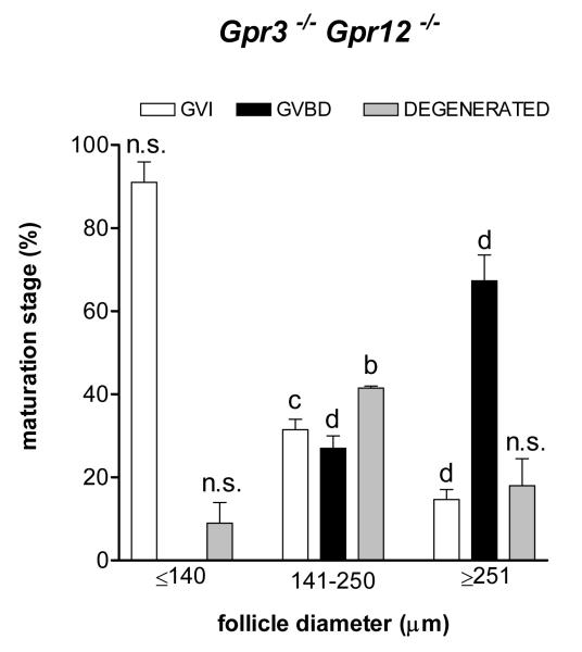
Although it is established that cAMP accumulation plays a pivotal role in preventing meiotic resumption in mammalian oocytes, the mechanisms controlling cAMP levels in the female gamete have remained elusive. Both production of cAMP via GPCRs/Gs/adenylyl cyclases endogenous to the oocyte as well as diffusion from the somatic compartment through gap junctions have been implicated in maintaining cAMP at levels that preclude maturation. Here we have used a genetic approach to investigate the different biochemical pathways contributing to cAMP accumulation and maturation in mouse oocytes. Because cAMP hydrolysis is greatly decreased and cAMP accumulates above a threshold, oocytes deficient in PDE3A do not resume meiosis in vitro or in vivo, resulting in complete female infertility. In vitro, inactivation of Gs or downregulation of the GPCR GPR3 causes meiotic resumption in the Pde3a null oocytes. Crossing of Pde3a(-/-) mice with Gpr3(-/-) mice causes partial recovery of female fertility. Unlike the complete meiotic block of the Pde3a null mice, oocyte maturation is restored in the double knockout, although it occurs prematurely as described for the Gpr3(-/-) mouse. The increase in cAMP that follows PDE3A ablation is not detected in double mutant oocytes, confirming that GPR3 functions upstream of PDE3A in the regulation of oocyte cAMP. Metabolic coupling between oocytes and granulosa cells was not affected in follicles from the single or double mutant mice, suggesting that diffusion of cAMP is not prevented. Finally, simultaneous ablation of GPR12, an additional receptor expressed in the oocyte, does not modify the Gpr3(-/-) phenotype. Taken together, these findings demonstrate that Gpr3 is epistatic to Pde3a and that fertility as well as meiotic arrest in the PDE3A-deficient oocyte is dependent on the activity of GPR3. These findings also suggest that cAMP diffusion through gap junctions or the activity of additional receptors is not sufficient by itself to maintain the meiotic arrest in the mouse oocyte.


Figures

Fig.1
Fig.1. Injection of Gs-neutralizing antibody or GPR3 morpholino oligonucleotides in Pde3a-/- null oocytes induces meiotic resumption
A. Groups of wild type (WT) oocytes maintained in meiotic arrest with 3.5 mM hypoxanthine or Pde3a-/- oocytes were injected with GPR3 morpholino oligonucleotide or a control scrambled morpholino as described (Hinckley et al., 2005). The effects of the injection on meiotic maturation were followed up to 24 hours. The data reported are the mean ± SE of the analysis from three experiments, and the meiotic maturation stage after 18 hours from the injections is reported as percentage of GVBD. The numbers at the top of the bars indicate the total number of oocytes injected. Superscripts indicate statistical difference vs. controls for each genotype: a, p<0.05; b, p<0.01; c, p<0.005 and d, p<0.001. B. Denuded oocytes from Pde3a-/- mice were injected with antibody against Gs or control rabbit IgG at a final concentration of 1.3 μM. Oocytes were monitored for meiotic resumption every 30 min for 3 hours and at 21-24 hours for PB extrusion. Meiotic maturation was scored after 2 hours from the injections and reported as percentage of GVBD. After 2 hours of culture, 96% of Gs antibody injected oocytes had resumed meiosis. The numbers above the bars indicate the total number of oocytes injected. The data are the average of two independent experiments.
Fig.2
Fig.2. Meiotic resumption in immature ovaries from mice with different genetic backgrounds
22-day-old mice were primed with 5 IU PMSG, ovaries were collected 42-44 hours later, and preovulatory follicles punctured to obtain COCs. Cumulus cells were stripped from oocytes with a pulled Pasteur pipette, and cell cycle progression immediately scored. Only well formed COCs were used for meiotic progression analysis. In all experiments M2 media supplemented with 3 mg/ml BSA was used. The bar graph represents mean ± SE of up to 4 different mice of the indicated genotype. A maximum of 15 min elapsed from the beginning of COC collection from each ovary to the end of the oocyte scoring. The letters above the bars indicate statistical difference within the same oocyte maturational stage, between the indicated genotype and Gpr3+/- Pde3a+/-. a, p<0.05; b, p<0.01; c, p<0.005 and d, p<0.001. n = number of oocytes scored for each genotype.
Fig.3
Fig.3. Ovarian morphology and state of oocyte maturation in Grp3-/- Pde3a-/- mice
22-day-old mice were primed with 5 IU PMSG; after 42-44 hours, ovaries were collected, fixed in 4% PFA and meiotic progression scored in follicles from HE sections. A. Median section of whole ovary from double mutant mice. B. Metaphase spindle in follicle enclosed oocytes from double null mice. C. Scoring of meiotic maturation in double heterozygous, Gpr3-/- Pde3a+/-, and Gpr3-/- Pde3a-/- mice. The state of oocyte maturation was related to the diameter of the follicle by measuring follicle size not including theca cells, as detailed in the methods. The data represent the mean ± SE of the analysis from three to five mice for each genotype. The letters above the bar indicate statistical difference between the oocyte maturation state in any given group of follicles of the indicated genotype and the corresponding maturation state follicles of the same diameter of Gpr3+/- Pde3a+/-. n.s., non significant; a, p<0.05; b, p<0.01; c, p<0.005 and d, p<0.001. No significant differences between Gpr3-/- and Gpr3-/- Pde3a-/- were observed.
Fig.4
Fig.4. State of oocyte maturation in Grp3-/- Gpr12-/- mice
The state of oocyte maturation was scored in Gpr3-/- Gpr12-/- and control mice. Every follicle and oocyte was analyzed using a 20X magnification. Follicle size, not including theca cells, was determined using the AxioVision 3.1.0.1.8 software (Carl Zeiss) and refers to the average of measurements in two dimensions. The graph represents the mean ± SE of the analysis from three mice for each genotype. The letters above the bar indicate statistical difference between the oocyte maturation state in any given group of follicles and the corresponding maturation state follicles of the same diameter of Gpr3+/- Pde3a+/-. n.s., non significant; a, p<0.05; b, p<0.01; c, p<0.005 and d, p<0.001. No significant differences among Grp3-/- Gpr12-/- and Gpr3-/- oocytes were observed.
Fig.5
Fig.5. cAMP levels in denuded oocytes from wild type and mice with ablation of Gpr3 and Pde3a
All oocytes analyzed were harvested from preantral follicles of 11- to12-day-old mice. Follicles isolated from ovaries were considered as preantral if smaller than 140 μm and without a visible antrum. Oocytes were collected without PDE inhibitors in M2 media supplemented with 3 mg/ml BSA. The collecting/denuding process was completed in less than 10 minutes/ovary. The graph reports mean ± SE of three to four different experiments. Numbers on the top of the bars indicate the total number of oocytes used. Different superscripts denote statistical difference between Gpr3+/+ Pde3a+/+ and the indicated genotype. a, p<0.05; b, p<0.01 and c, p<0.005.
Fig. 6
Fig. 6. Gap junction permeability is intact in double mutant mice
Metabolic coupling between oocyte and granulosa cells was measured in preantral follicles. Follicles isolated from 11- to 12-day-old mice were incubated in M2/BSA supplemented with 0.1% type I collagenase and 0.01 U/ul DNaseI for 20 min at 37°C. Half of the oocyte-granulosa cell complexes were used to obtain denuded oocytes, and then the two groups were incubated for 1 hour in the presence of [3H] uridine (10μCi/group). The difference of uptake between the groups reflects the transport of [3H] uridine through gap junctions. The graph shown represents the mean ± SE of three independent experiments using quadruplicates for each group of oocytes. The numbers at the top of the bars indicate the total number of oocytes tested. No significant differences between Gpr3+/- Pde3a+/- and Gpr3-/- Pde3a+/- or Gpr3-/- Pde3a-/- were observed. N refers to the final number of animals used for each genotype.

References

    1. Aberdam E, Hanski E, Dekel N. Maintenance of meiotic arrest in isolated rat oocytes by the invasive adenylate cyclase of Bordetella pertussis. Biol Reprod. 1987;36:530–5. - PubMed
    1. Ackert CL, Gittens JE, O’Brien MJ, Eppig JJ, Kidder GM. Intercellular communication via connexin43 gap junctions is required for ovarian folliculogenesis in the mouse. Dev Biol. 2001;233:258–70. - PubMed
    1. Anderson E, Albertini DF. Gap junctions between the oocyte and companion follicle cells in the mammalian ovary. J Cell Biol. 1976;71:680–6. - PMC - PubMed
    1. Bornslaeger EA, Schultz RM. Regulation of mouse oocyte maturation: effect of elevating cumulus cell cAMP on oocyte cAMP levels. Biol Reprod. 1985;33:698–704. - PubMed
    1. Chesnel F, Wigglesworth K, Eppig JJ. Acquisition of meiotic competence by denuded mouse oocytes: participation of somatic-cell product(s) and cAMP. Dev Biol. 1994;161:285–95. - PubMed

Publication types

MeSH terms

LinkOut - more resources