

Control of ion selectivity in LeuT: two Na+ binding sites with two different mechanisms
- PMID: 18280500
- PMCID: PMC4948944
- DOI: 10.1016/j.jmb.2008.01.015
Control of ion selectivity in LeuT: two Na+ binding sites with two different mechanisms
Abstract
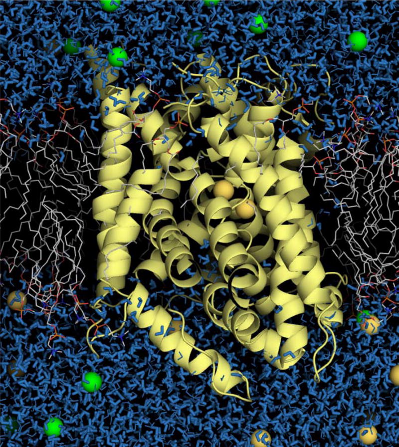
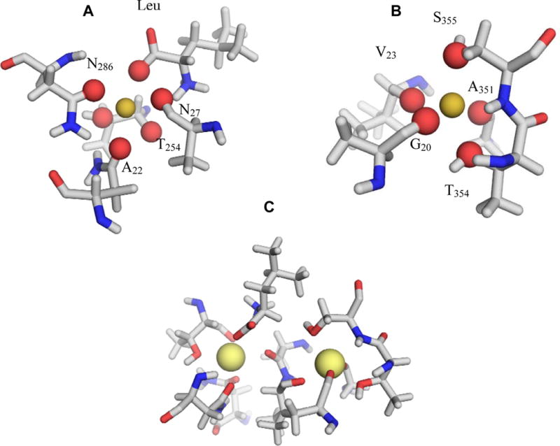
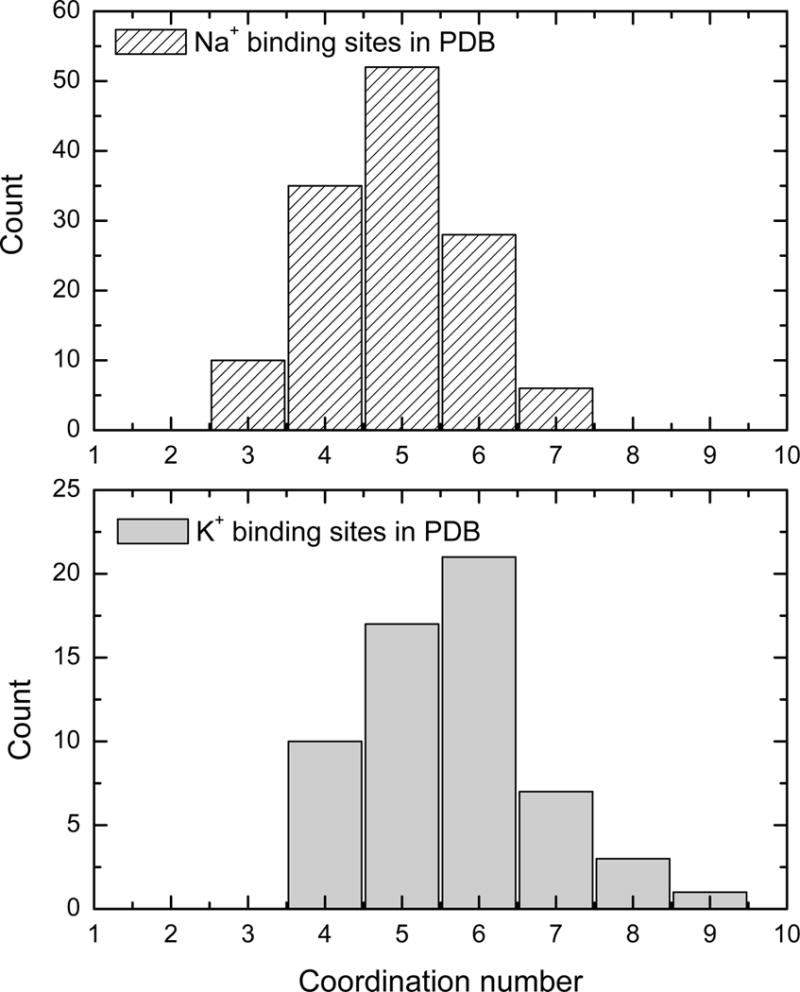
The x-ray structure of LeuT, a bacterial homologue of Na(+)/Cl(-)-dependent neurotransmitter transporters, provides a great opportunity to better understand the molecular basis of monovalent cation selectivity in ion-coupled transporters. LeuT possesses two ion binding sites, NA1 and NA2, which are highly selective for Na(+). Extensive all-atom free-energy molecular dynamics simulations of LeuT embedded in an explicit membrane are performed at different temperatures and various occupancy states of the binding sites to dissect the molecular mechanism of ion selectivity. The results show that the two binding sites display robust selectivity for Na(+) over K(+) or Li(+), the competing ions of most similar radii. Of particular interest, the mechanism primarily responsible for selectivity for each of the two binding sites appears to be different. In NA1, selectivity for Na(+) over K(+) arises predominantly from the strong electrostatic field arising from the negatively charged carboxylate group of the leucine substrate coordinating the ion directly. In NA2, which comprises only neutral ligands, selectivity for Na(+) is enforced by the local structural restraints arising from the hydrogen-bonding network and the covalent connectivity of the polypeptide chain surrounding the ion according to a "snug-fit" mechanism.
Figures


Similar articles
-
Molecular mechanism of ion-ion and ion-substrate coupling in the Na+-dependent leucine transporter LeuT.Biophys J. 2008 Nov 15;95(10):4613-21. doi: 10.1529/biophysj.108.139741. Epub 2008 Aug 15. Biophys J. 2008. PMID: 18708457 Free PMC article.
-
Conformational Dynamics on the Extracellular Side of LeuT Controlled by Na+ and K+ Ions and the Protonation State of Glu290.J Biol Chem. 2016 Sep 16;291(38):19786-99. doi: 10.1074/jbc.M116.731455. Epub 2016 Jul 29. J Biol Chem. 2016. PMID: 27474737 Free PMC article.
-
Molecular dynamics simulations of Na(+) and leucine transport by LeuT.Biochem Biophys Res Commun. 2015 Aug 14;464(1):281-5. doi: 10.1016/j.bbrc.2015.06.143. Epub 2015 Jun 24. Biochem Biophys Res Commun. 2015. PMID: 26116773
-
How LeuT shapes our understanding of the mechanisms of sodium-coupled neurotransmitter transporters.J Physiol. 2014 Mar 1;592(5):863-9. doi: 10.1113/jphysiol.2013.259051. Epub 2013 Jul 22. J Physiol. 2014. PMID: 23878376 Free PMC article. Review.
-
Neurotransmitter transporters: structure meets function.Structure. 2013 May 7;21(5):694-705. doi: 10.1016/j.str.2013.03.002. Structure. 2013. PMID: 23664361 Free PMC article. Review.
Cited by
-
A molecular switch controls the impact of cholesterol on a Kir channel.Proc Natl Acad Sci U S A. 2022 Mar 29;119(13):e2109431119. doi: 10.1073/pnas.2109431119. Epub 2022 Mar 25. Proc Natl Acad Sci U S A. 2022. PMID: 35333652 Free PMC article.
-
Exploring the ion selectivity properties of a large number of simplified binding site models.Biophys J. 2010 Jun 16;98(12):2877-85. doi: 10.1016/j.bpj.2010.03.038. Biophys J. 2010. PMID: 20550900 Free PMC article.
-
Conformational rearrangements to the intracellular open states of the LeuT and ApcT transporters are modulated by common mechanisms.Biophys J. 2010 Dec 15;99(12):L103-5. doi: 10.1016/j.bpj.2010.10.003. Biophys J. 2010. PMID: 21156121 Free PMC article.
-
Structural Mechanism of ω-Currents in a Mutated Kv7.2 Voltage Sensor Domain from Molecular Dynamics Simulations.J Chem Inf Model. 2021 Mar 22;61(3):1354-1367. doi: 10.1021/acs.jcim.0c01407. Epub 2021 Feb 11. J Chem Inf Model. 2021. PMID: 33570938 Free PMC article.
-
Exploring the Structural Dynamics of LeuT Using EPR Spectroscopy: A Focus on Transmembrane Helix 10.J Neurochem. 2025 Mar;169(3):e70034. doi: 10.1111/jnc.70034. J Neurochem. 2025. PMID: 40052253 Free PMC article.
References
-
- Hille B. Ion Channels of Excitable Membranes. 3rd. Sinauer; Sunderland, MA: 2001.
-
- DeFelice L. Transporter structure and mechanism. Trends Neurosci. 2004;27:352–359. - PubMed
-
- Rudnick G. In: Neurotransmitter transporters: Structure, Functions and Regulation. Reith RA, editor. Humana Press; Totowa, NJ, USA: 2002.
Publication types
MeSH terms
Substances
Grants and funding
LinkOut - more resources
Full Text Sources
Other Literature Sources
