. 2008 Apr 25;283(17):11526-34.
doi: 10.1074/jbc.M708935200. Epub 2008 Feb 14.

The tec family tyrosine kinase Btk Regulates RANKL-induced osteoclast maturation

Affiliations

The tec family tyrosine kinase Btk Regulates RANKL-induced osteoclast maturation

Seoung Hoon Lee et al. J Biol Chem. .

Abstract

A spontaneous mutation in Bruton's tyrosine kinase (Btk) induces a defect in B-cell development that results in the immunodeficiency diseases X-linked agammaglobulinemia in humans and X-linked immunodeficiency (Xid) in mice. Here we show an unexpected role of Btk in osteoclast formation. When bone marrow cells derived from Xid mice were stimulated with receptor activator of NF-kappaB ligand, an osteoclast differentiation factor, they did not completely differentiate into mature multinucleated osteoclasts. Moreover, we found that the defects appeared to occur at the stage in which mononuclear preosteoclasts fuse to generate multinucleated cells. Supporting this notion, macrophages from Xid mice also failed to form multinucleated foreign body giant cells. The fusion defect of the Xid mutant osteoclasts was caused by decreased expression of nuclear factor of activated T cells c1 (NFATc1), a master regulator of osteoclast differentiation, as well as reduced expression of various osteoclast fusion-related molecules, such as the d2 isoform of vacuolar H(+)-ATPase V0 domain and the dendritic cell-specific transmembrane protein. This deficiency was completely rescued by the introduction of a constitutively active form of NFATc1 into bone marrow-derived macrophages. Our data provide strong evidence that Btk plays a critical role in osteoclast multinucleation by modulating the activity of NFATc1.


Figures

FIGURE 1.
FIGURE 1.
Decreased osteoclast formation from the bone marrow cells of Xid mice in a coculture system. A, induction of Btk expression during osteoclastogenesis. BMMs from wild-type mice were cultured with M-CSF and RANKL for 4 days. Total lysates were subjected to SDS-PAGE and Western blot analysis to detect Btk. B and C, osteoclast formation in a coculture system. Bone marrow cells derived from wild-type (WT) and Xid mice were cultured with osteoblasts in the presence of 1α, 25(OH)2D3 and PGE2 for 9 days. B, cells were fixed and stained for TRAP. C, TRAP(+) MNCs with more than three nuclei (left panel) or more than three nuclei and larger than 100 μm in diameter (right panel) were counted as osteoclasts. D, a TRAP solution assay. TRAP activity was assessed at an absorbance of 405 nm. Data are expressed as the mean ± S.D. and are representative of at least three experiments. NS, not significant. *, p < 0.05 versus WT; †, p < 0.005 versus WT. Scale bar, 200 μm.
FIGURE 2.
FIGURE 2.
Decreased TRAP(+) MNCs and pit formation by bone marrow cells derived from Xid mice in stromal cell-free BMM cultures. A and B, osteoclast formation in BMM cultures. BMMs were cultured with 30 ng/ml M-CSF and RANKL at the indicated concentrations for 4 days. A, cells were fixed and stained with rhodamine-phalloidin to label the F-actin ring, followed by TRAP staining. B, TRAP(+) MNCs with more than three nuclei (left panel) or more than three nuclei and larger than 100 μm in diameter (right panel) were counted as osteoclasts. C, the TRAP solution assay was performed as described under “Experimental Procedures.” D, pit formation. BMMs were cultured on dentine slices with M-CSF and RANKL for 4 days. Resorption pits were stained with 0.5% toluidine blue and counted. Data are expressed as the mean ± S.D. and are representative of at least three experiments. *, p < 0.05 versus WT; †, p < 0.01 versus WT. Scale bar, 200 μm.
FIGURE 3.
FIGURE 3.
Impaired fusion of preosteoclasts from Xid mice. A, the formation of TRAP(+) mononuclear preosteoclasts. The formation of preosteoclasts (pOCs) was induced by coculturing bone marrow cells derived from wild-type (WT) or Xid mice with osteoblasts in the presence of 1α, 25(OH)2D3 and PGE2 for 6 days. Cells were stained for TRAP (left panel), and TRAP(+) mononuclear cells were counted as preosteoclasts (right panel). B, mature osteoclast formation from preosteoclasts. Preosteoclasts obtained as described in A were differentiated into osteoclasts in the presence of M-CSF and RANKL for 24 h. Cells were fixed and stained for TRAP (left panel). TRAP(+) MNCs with more than three nuclei (upper right panel) or more than three nuclei and larger than 100 μm in diameter (lower right panel) were counted as fused osteoclasts. C, a fusion efficiency assay. The efficiency of preosteoclast fusion was calculated by dividing the total number of nuclei stained with 4′,6-diamidino-2-phenylindole within TRAP(+) MNCs by the number of TRAP(+) MNCs. D, the formation of foreign body giant cells (FBGCs). BMMs from wild-type and Xid mice were treated with IL-3 and IL-4 to induce giant cell formation. Cells were stained with May-Grünwald-Giemsa (left). Giant cells (>100 μm in diameter) were counted (right). Data are represented as the mean ± S.D. of experiments performed in triplicate. NS, not significant. *, p < 0.01 versus WT; †, p < 0.001 versus WT. Scale bar, 200 μm.
FIGURE 4.
FIGURE 4.
Decreased expression and nuclear translocation of NFATc1 in preosteoclasts from Xid. A, NFATc1 mRNA levels during osteoclast differentiation in BMM cultures. BMMs from wild-type (WT) and Xid mice were cultured with M-CSF and RANKL for 4 days. RNA was isolated at the indicated time points and subjected to real-time quantitative PCRs to analyze the levels of NFATc1 mRNA. B, NFATc1 mRNA levels during preosteoclast fusion. To generate preosteoclasts, bone marrow cells from wild-type (WT) and Xid mice were cultured with osteoblasts in the presence of 1α, 25(OH)2D3 and PGE2 for 6 days. Preosteoclasts were treated with M-CSF and RANKL to induce cell-cell fusion. RNA was isolated at the indicated time points and subjected to real-time quantitative PCRs as described in A. C and D, NFATc1 expression and translocation during preosteoclast fusion. Preosteoclasts obtained as described in B were induced to fuse for 12 h with M-CSF alone or M-CSF plus RANKL in the absence or presence of CsA (1 μg/ml). Total lysates (C) or cytosolic and nuclear fractions (D) were harvested from cultured cells and subjected to SDS-PAGE and Western blot analysis to detect NFATc1. Antibodies specific for actin and histone H3 were used to normalize the cytosolic and nuclear extracts, respectively. E, PLCγ2 activation by RANKL in WT and Xid mutant cells. BMMs from WT and Xid mice were stimulated with RANKL (100 ng/ml) after 6 h of starvation in serum-free medium. Total lysates were subjected to SDS-PAGE and Western blot analysis. Data are expressed as the mean ± S.D. and are representative of three independent experiments. F, NFATc1 mRNA levels were measured as described in A. Cells were treated with RANKL for 24 h in the absence or presence of CsA (1 μg/ml). *, p < 0.05; NS, not significant.
FIGURE 5.
FIGURE 5.
Expression levels of fusion-related molecules during the differentiation of BMMs derived from Xid mice into osteoclasts. BMMs from wild-type (WT) and Xid mice were cultured with M-CSF and RANKL for 4 days. RNA was isolated on the indicated days and subjected to real-time quantitative PCR analysis. Day 0 indicates that the cells were BMMs. A, levels of mRNA encoding osteoclast-specific markers, including OSCAR, TRAP, and cathepsin K. B, levels of mRNA encoding fusion-related (Atp6v0d2 and DC-STAMP) and adhesion molecules (Integrin αv and Integrin β3). Data are expressed as the mean ± S.D. and are representative of three independent experiments.
FIGURE 6.
FIGURE 6.
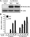
Restoration of osteoclast formation by ectopic expression of NFATc1 in BMMs derived from Xid mice. A, overexpression of NFATc1. BMMs from wild-type (WT) and Xid mice were infected with pMX-puro (control retrovirus) or retrovirus encoding HA-tagged Ca-NFATc1 (constitutively active form) and selected with puromycin (2 μg/ml) for 48 h. Retroviral-mediated expression of Ca-NFATc1 was analyzed by Western blotting with antibodies specific for HA. B, effects of NFATc1 overexpression on osteoclast differentiation. BMMs infected with retrovirus were differentiated into osteoclasts in the presence of M-CSF (30 ng/ml) and RANKL (0–200 ng/ml) for 4 days. Cultured cells were fixed and stained for TRAP. TRAP(+) MNCs with more than three nuclei and larger than 100 μm in diameter were counted as osteoclasts. Data are expressed as the mean ± S.D. and are representative of at least three experiments. *, p < 0.02 versus WT; †, p < 0.001 versus WT.
FIGURE 7.
FIGURE 7.
A schematic model of the induction of NFATc1 activation during RANKL-stimulated osteoclastogenesis. ITAM activation resulting from stimulation via immunoreceptors (OSCAR and TREM-2) or the RANKL-RANK interaction may lead to activation of Btk and subsequent recruitment of downstream effectors, including PLCγ2. PLCγ2 activation induces calcium signaling, which is critical for NFATc1 activation. Alternatively, Btk may recruit and activate undefined effector molecules that, in turn, mediate NFATc1 activation. NFATc1 activation is also dependent on c-Fos and TRAF6, both of which are activated by RANKL. Finally, activated NFATc1 may contribute to osteoclast fusion, differentiation, and activation.

References

    1. Walsh, M. C., Kim, N., Kadono, Y., Rho, J., Lee, S. Y., Lorenzo, J., and Choi, Y. (2006) Annu. Rev. Immunol. 24 33-63 - PubMed
    1. Teitelbaum, S. L. (2000) Science 289 1504-1508 - PubMed
    1. Boyle, W. J., Simonet, W. S., and Lacey, D. L. (2003) Nature 423 337-342 - PubMed
    1. Lee, S. H., Rho, J., Jeong, D., Sul, J. Y., Kim, T., Kim, N., Kang, J. S., Miyamoto, T., Suda, T., Lee, S. K., Pignolo, R. J., Koczon-Jaremko, B., Lorenzo, J., and Choi, Y. (2006) Nat. Med. 12 1403-1409 - PubMed
    1. Yagi, M., Miyamoto, T., Sawatani, Y., Iwamoto, K., Hosogane, N., Fujita, N., Morita, K., Ninomiya, K., Suzuki, T., Miyamoto, K., Oike, Y., Takeya, M., Toyama, Y., and Suda, T. (2005) J. Exp. Med. 202 345-351 - PMC - PubMed

Publication types

LinkOut - more resources