. 2008 Apr 18;283(16):10276-86.
doi: 10.1074/jbc.M801160200. Epub 2008 Feb 15.

Identification of FRA1 and FRA2 as genes involved in regulating the yeast iron regulon in response to decreased mitochondrial iron-sulfur cluster synthesis

Affiliations

Identification of FRA1 and FRA2 as genes involved in regulating the yeast iron regulon in response to decreased mitochondrial iron-sulfur cluster synthesis

Attila Kumánovics et al. J Biol Chem. .

Abstract

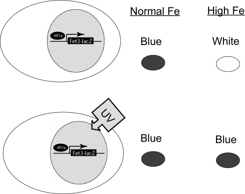
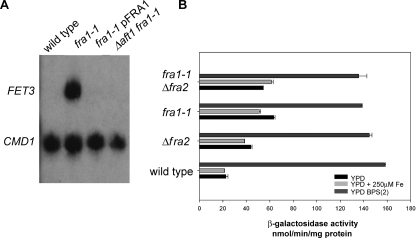
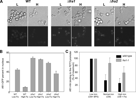
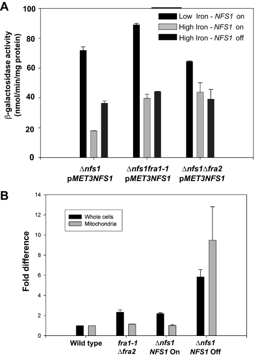
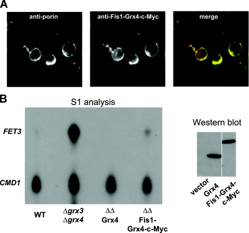
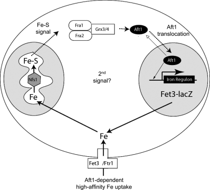
The nature of the connection between mitochondrial Fe-S cluster synthesis and the iron-sensitive transcription factor Aft1 in regulating the expression of the iron transport system in Saccharomyces cerevisiae is not known. Using a genetic screen, we identified two novel cytosolic proteins, Fra1 and Fra2, that are part of a complex that interprets the signal derived from mitochondrial Fe-S synthesis. We found that mutations in FRA1 (YLL029W) and FRA2 (YGL220W) led to an increase in transcription of the iron regulon. In cells incubated in high iron medium, deletion of either FRA gene results in the translocation of the low iron-sensing transcription factor Aft1 into the nucleus, where it occupies the FET3 promoter. Deletion of either FRA gene has the same effect on transcription as deletion of both genes and is not additive with activation of the iron regulon due to loss of mitochondrial Fe-S cluster synthesis. These observations suggest that the FRA proteins are in the same signal transduction pathway as Fe-S cluster synthesis. We show that Fra1 and Fra2 interact in the cytosol in an iron-independent fashion. The Fra1-Fra2 complex binds to Grx3 and Grx4, two cytosolic monothiol glutaredoxins, in an iron-independent fashion. These results show that the Fra-Grx complex is an intermediate between the production of mitochondrial Fe-S clusters and transcription of the iron regulon.


Figures

FIGURE 1.
FIGURE 1.
Model of the genetic screen designed to identify genes that regulate the iron regulon. Yeast cells containing a FET3-lacZ reporter gene integrated at the HO locus were deleted for the FET3 gene. When grown in standard culture medium, these cells will express FET3-lacZ due to impaired iron transport, thereby turning blue when exposed to X-gal, a substrate for β-galactosidase. The addition of 50 μm iron to the growth medium will completely suppress FET3-lacZ expression, since cells obtain iron through low affinity iron transporters. Based on this observation, wild type cells containing the integrated FET3-lacZ reporter were mutagenized by UV and plated on medium supplemented with 50 μm iron. Colonies that were blue in iron-replete medium were further studied.
FIGURE 2.
FIGURE 2.
Deletion or mutation of FRA genes affects transcription of FET3. A, wild type, fra1-1, and fra1-1 cells transformed with a plasmid expressing FRA1 under the control of the endogenous promoter and fra1-1Δaft1 cells were incubated in high iron medium (YPD + 250 μm FeSO4). Cells were grown to midlog phase and harvested, and mRNA levels for FET3 and for calmodulin (CMD1) were analyzed by S1 analysis. B, wild type, fra1-1, Δfra2, and fra1-1Δfra2 cells containing an integrated FET3-lacZ reporter construct were incubated in low iron (dark gray bars; YPD + 40 μm BPS + 2 μm FeSO4), iron-sufficient (black bars; YPD), and high iron (light gray bars; YPD + 250μm FeSO4) medium. The cells were grown for 12 h, and β-galactosidase activity was determined.
FIGURE 3.
FIGURE 3.
Aft1 enters the nucleus and occupies the FET3 promoter in Δfra1 cells. A, wild type (WT), Δfra1, and Δfra2 cells were transformed with a low copy plasmid expressing a GFP-tagged Aft1 under the control of its endogenous promoter. The cells were grown overnight in low (L) and high iron (H) medium, and the subcellular localization of Aft1-GFP was examined by fluorescence microscopy. B, the percentage of cells showing Aft1-GFP in the nucleus was quantified. C, wild type (black bars) and fra1-1 cells (gray bars) were transformed with a low copy plasmid containing AFT1-HA. The cells were grown in iron-limited (CM + 40 μm BPS iron), “normal” iron (CM), or iron-replete (CM + 250 μm Fe2SO4) media. After 12 h of growth, cells were harvested, and chromatin was immunoprecipitated using the HA epitope of Aft1. After cross-linking was reversed, PCR was performed using primers, which flank the Aft1 binding site of the FET3 promoter, with CMD1 used as a positive control. Aft1 binding to the FET3 promoter in normal and high iron conditions is shown as the percentage of the low iron (maximum) values, and the error bars represent the S.E. These results are the average of three independent experiments.
FIGURE 4.
FIGURE 4.
Fra1 and Fra2 are part of the mitochondrial Fe-S cluster synthesis-sensing pathway. A, cells (Δnfs1, Δnfs1 fra1-1, Δnfs1Δfra2) containing an integrated FET3-lacZ reporter construct were transformed with a plasmid containing MET3-regulated NFS1. Cells were grown in medium lacking methionine to induce expression of NFS1. The cells were transferred to medium lacking methionine (on) or medium containing 10 times the normal level of methionine (off) to prevent expression of NFS1. The media were either iron-limited (black bars; low iron CM + 40 μm BPS) or iron-replete (high iron CM + 50 μm FeSO4). The cells were grown in the specified media for 6 h, and then β-galactosidase activity was measured. B, wild type, fra1-1Δfra2, and Δnfs1(pMET3NFS1) cells were grown in CM medium in the presence or absence of methionine for 12 h. Cells were harvested, and the amounts of iron in whole cells and purified mitochondria were determined by inductively coupled plasma mass spectrometry. The data were normalized to cell or mitochondrial protein, and μg of iron/mg of protein is expressed as -fold difference compared with wild type levels. The error bars are the S.D. of three experiments.
FIGURE 5.
FIGURE 5.
Depletion of Fra1 does not affect mitochondrial aconitase. A, fra1-1 cells containing a FET3-lacZ reporter construct were transformed with a plasmid containing a β-estradiol-inducible FRA1. The cells were grown in CM in the presence of β-estradiol, and then either cells were maintained with β-estradiol, or β-estradiol was removed for the specified times. Samples were taken, and Fra1-His6 protein was determined by Western blot analysis. Aliquots were also taken to assay β-galactosidase (B) and aconitase (C) activity.
FIGURE 6.
FIGURE 6.
Depletion of Nfs1 affects aconitase and induces expression of FET3-lacZ. A, Δnfs1 cells containing pMET3NFS1 and a FET3-lacZ reporter were grown for 8 h in normal (CM), high iron (50 μm FeSO4), or low iron media (CM + 40 μm BPS + 2 μm FeSO4) either in the presence (black bars) or absence of methionine (gray bar). The cells were harvested, and aconitase (A) and β-galactosidase (B) activity was measured.
FIGURE 7.
FIGURE 7.
Immunoprecipitation of Fra2-His6 protein identifies a protein complex. A, cells (Δfra2) were transformed with a low copy plasmid containing a GRX4-c-Myc construct and a plasmid containing FRA2-His6 or FRA2 without the epitope tag. The cells were grown for 6 h in either low (L; CM + 40 μm BPS) or in high iron (H; CM + 50 μm FeSO4) medium. Extracts were applied to Ni2+-NTA resin, and the eluates were tested for the indicated proteins by Western blotting. Grx4-c-Myc was detected using an antibody specific for c-Myc. Fra2-His6 was detected using an anti-His antibody, and Fra1 was detected using a rabbit anti-Fra1 antibody. B, cells (Δfra2) were transformed with a high copy plasmid containing FRA2-His6 or FRA2 without the epitope tag. Cells were grown as in A and affinity-purified as in A, and eluates were probed for Fra2-His6 and endogenous Grx3 using anti-His6 or rabbit anti-Grx3 antibodies. C, wild type (WT) cells were grown as in A, extracts were obtained, and Fra2 was immunopurified using rabbit anti-Fra2 antibodies bound to Dynabeads (sheep anti-rabbit IgG; Invitrogen). Samples were tested for Grx3 and Fra2 by Western blotting. D, wild type cells were transformed with plasmids containing Grx4-YFP and Fra2-YFP. Cells were grown on CM-Ura-Leu supplemented with either FeS04 (100 μm) or BPS (100 μm) at 30 °C. After 20 h, cells were diluted 100× into fresh media containing doxycycline (1 μg/ml). After 18 h, the cultures were serially diluted (50× and then 20×) into fresh media and grown for an additional 20 h. Aliquots were removed from both dilutions and treatments, stained with 4′,6-diamidino-2-phenylindole (DAPI), and fixed to microscope slides treated with concanavalin A. Cells were stained with 4′,6-diamidino-2-phenylindole, mounted onto a coverslip, and examined for YFP and 4′,6-diamidino-2-phenylindole fluorescence, using an Olympus BX51 microscope with a ×100 1.4A oil immersion lens. Images were captured using PictureFramer software. DIC, differential interference contrast.
FIGURE 8.
FIGURE 8.
Aft1-TAP co-immunoprecipitates with Grx3. Cells containing an AFT1-TAP integrated into the genome were grown for 6 h in high and low iron medium. Cells were lysed and immunoprecipitated as described under “Experimental Procedures” using a Grx3-specific antibody. The immunoprecipitate (IP) was probed for Aft1-TAP using a polyclonal anti-TAP antibody and a polyclonal anti-Grx3 using Western blot analysis.
FIGURE 9.
FIGURE 9.
Grx3 localizes to the cytoplasm. A, wild type, Δgrx3, or wild type (WT) cells transformed with a high copy plasmid containing GRX3 under the control of its endogenous promoter, were grown in high (H) and low (L) iron media and processed for immunofluorescence using a polyclonal anti-Grx3 antibody (1:100) and the monoclonal anti-Pgk1 antibody followed by Alexa 594-conjugated goat anti-rabbit IgG and Alexa 488-conjugated goat anti-mouse IgG (1:750; Invitrogen). DIC, differential interference contrast. B, wild type cells were grown to midlog phase and homogenized, and postnuclear supernatant was separated into a postmitochondrial fraction (PMS) and a crude mitochondrial fraction (Crude mito). Mitochondria were further purified by ultracentrifugation in a sucrose gradient (post gradient). Equal amounts of protein are loaded in each lane. The presence of organellar markers was determined by Western blot using anti-Grx3, anti-Pgk1 (cytosolic marker), anti-CPY (vacuolar marker), anti-porin (mitochondrial marker), and anti-Dpm1 (endoplasmic reticulum ER marker) (Invitrogen).
FIGURE 10.
FIGURE 10.
Cytosolic Grx3-Grx4 affects Aft1 function. A, cells expressing Fis1-Grx4-c-Myc fusion protein were grown to midlog and processed for immunofluorescence using rabbit anti-c-Myc (1:100; Covance) and mouse anti-porin (1:100; Invitrogen), followed by Alexa 594-conjugated goat anti-rabbit IgG and Alexa 488-conjugated goat anti-mouse IgG (1:750; Invitrogen). The c-Myc-tagged Fis1-Grx4 co-localized with the mitochondrial outer membrane protein porin, showing that the fusion protein is tethered to the mitochondria, thus restricting Grx4 to the cytosol. B, wild type (WT), Δgrx3Δgrx4, or Δgrx3Δgrx4 transformed with empty vector, pGRX4-c-Myc, or pFIS1-GRX4-c-Myc were grown to midlog, and samples were harvested for FET3 and CMD1 S1 analysis or Western blot using an anti-c-Myc tag antibody. Grx4-c-Myc and Fis1-Grx4-c-Myc are expressed at comparable levels.
FIGURE 11.
FIGURE 11.
Model for the role of Fra1-Fra2 in the regulation of Aft1 transcriptional activity. In the absence of mitochondrial Fe-S cluster synthesis, Aft1 translocates into the nucleus and induces the transcription of its target genes, such as the plasma membrane high affinity iron permease complex Fet3-Ftr1. The signal from the mitochondria is not known, but it is interpreted in the cytosol by a protein complex consisting of the Fra1-Fra2 proteins with Grx3-Grx4. This complex inhibits the translocation of Aft1 into the nucleus. Our data suggest that Aft1 does not bind to the Fra1-Fra2 complex, indicating that there is not a direct physical interaction between the two. The Fra1-Fra2-Grx complex may affect sulfhydryl status on Aft1 or on an Aft1-interacting protein. Low iron conditions reduce the rate of Fe-S cluster synthesis, resulting in a signal through the Fra1-Fra2-Grx complex and in Aft1 translocating to the nucleus, where it occupies the FET3 promoter. Further decreases in cytosolic iron may affect the amount of Aft1 bound to the promoter, leading to an increase in transcriptional activation.

References

    1. Van Ho, A., Ward, D. M., and Kaplan, J. (2002) Annu. Rev. Microbiol. 56 237–261 - PubMed
    1. Yamaguchi-Iwai, Y., Dancis, A., and Klausner, R. D. (1995) EMBO J. 14 1231–1239 - PMC - PubMed
    1. Yamaguchi-Iwai, Y., Ueta, R., Fukunaka, A., and Sasaki, R. (2002) J. Biol. Chem. 277 18914–18919 - PubMed
    1. Blaiseau, P. L., Lesuisse, E., and Camadro, J. M. (2001) J. Biol. Chem. 276 34221–34226 - PubMed
    1. Rutherford, J. C., Jaron, S., Ray, E., Brown, P. O., and Winge, D. R. (2001) Proc. Natl. Acad. Sci. U. S. A. 98 14322–14327 - PMC - PubMed

Publication types

MeSH terms