

Quantitative second harmonic generation imaging of the diseased state osteogenesis imperfecta: experiment and simulation
- PMID: 18281387
- PMCID: PMC2480682
- DOI: 10.1529/biophysj.107.114405
Quantitative second harmonic generation imaging of the diseased state osteogenesis imperfecta: experiment and simulation
Abstract
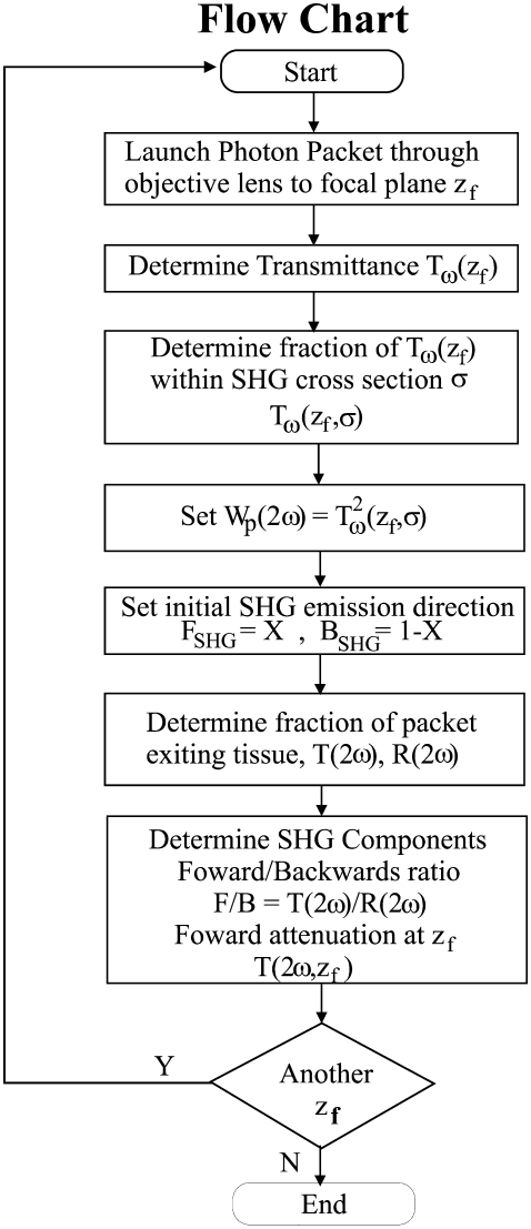
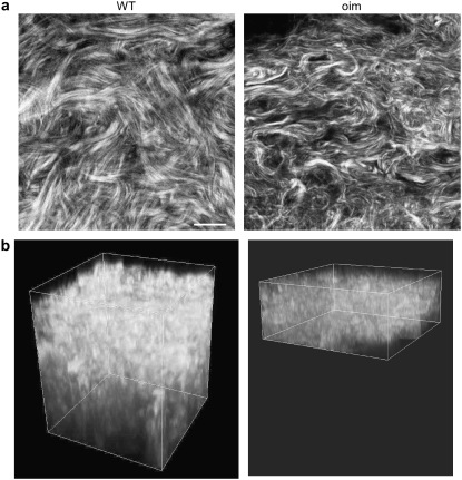
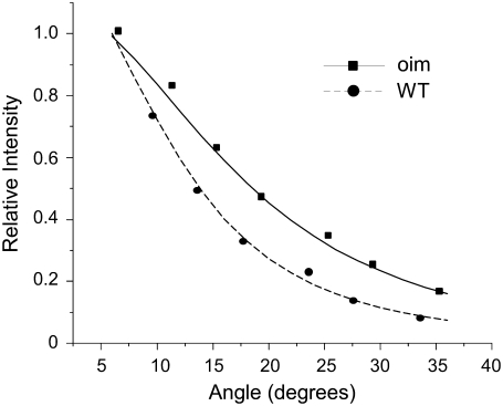
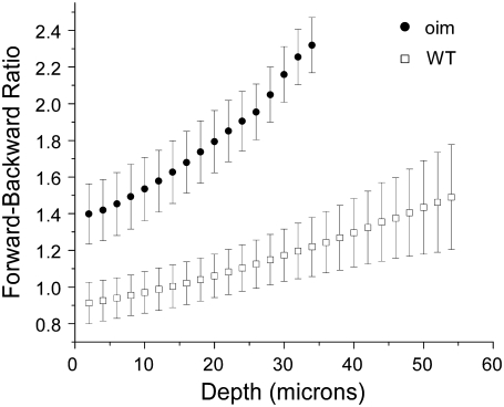
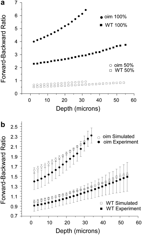
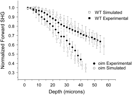
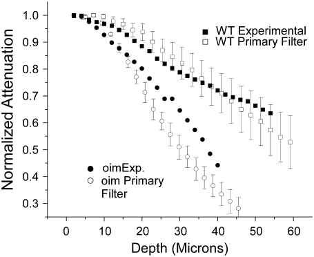
We report the integrated use of 3D second harmonic generation (SHG) imaging microscopy and Monte Carlo simulation as a combined metric to quantifiably differentiate normal and diseased tissues based on the physical properties of the respective extracellular matrix. To achieve this, we have identified a set of parameters comprised of the SHG creation attributes and the bulk optical parameters, which are used collectively via comparative analysis. Monte Carlo simulations of the SHG axial directional and attenuation responses allow their decomposition into the underlying factors that are not readily obtainable through experimental techniques. Specifically, this approach allows for estimation of the SHG creation attributes (directionality and relative conversion efficiency) and separation of primary and secondary filter effects, collectively that form the observed SHG contrast. The quantitative metric is shown for the connective tissue disorder Osteogenesis Imperfecta (characterized by abnormal assembly of type I collagen) using a murine model that expresses the disease in the dermis layer of skin. Structural dissimilarities between the osteogenesis imperfecta mouse and wild-type tissues lead to significant differences in the SHG depth-dependent directionality and signal attenuation. The Monte Carlo simulations of these responses using measured bulk optical parameters reproduce the experimental data trends, and the extracted emission directionality and conversion efficiencies are consistent with independent determinations. The simulations also illustrate the dominance of primary filter affects on overall SHG generation and attenuation. Thus, the combined method of 3D SHG imaging and modeling forms an essential foundation for parametric description of the matrix properties that are not distinguishable by sole consideration of either bulk optical parameters or SHG alone. Moreover, due to the quasi-coherence of the SHG process in tissues, we submit that this approach contains unique information not possible by purely scattering based methods and that these methods will be applicable in the general case where the complex fibrillar structure is difficult to fully quantify via morphological analysis.
Figures
Similar articles
-
Second harmonic generation imaging microscopy studies of osteogenesis imperfecta.J Biomed Opt. 2007 Sep-Oct;12(5):051805. doi: 10.1117/1.2799538. J Biomed Opt. 2007. PMID: 17994883
-
Alterations of the extracellular matrix in ovarian cancer studied by Second Harmonic Generation imaging microscopy.BMC Cancer. 2010 Mar 11;10:94. doi: 10.1186/1471-2407-10-94. BMC Cancer. 2010. PMID: 20222963 Free PMC article.
-
Second-harmonic generation circular dichroism studies of osteogenesis imperfecta.Opt Lett. 2012 Sep 15;37(18):3837-9. doi: 10.1364/ol.37.003837. Opt Lett. 2012. PMID: 23041876 Free PMC article.
-
From molecular structure to tissue architecture: collagen organization probed by SHG microscopy.J Biophotonics. 2013 Feb;6(2):129-42. doi: 10.1002/jbio.201200092. Epub 2012 Jul 12. J Biophotonics. 2013. PMID: 22791562 Review.
-
Second-harmonic generation imaging of cancer.Methods Cell Biol. 2014;123:531-46. doi: 10.1016/B978-0-12-420138-5.00028-8. Methods Cell Biol. 2014. PMID: 24974046 Review.
Cited by
-
Collagen organization of renal cell carcinoma differs between low and high grade tumors.BMC Cancer. 2019 May 23;19(1):490. doi: 10.1186/s12885-019-5708-z. BMC Cancer. 2019. PMID: 31122202 Free PMC article.
-
Application of quantitative second-harmonic generation microscopy to dynamic conditions.Biomed Opt Express. 2013 Oct 21;4(11):2546-54. doi: 10.1364/BOE.4.002546. eCollection 2013. Biomed Opt Express. 2013. PMID: 24298415 Free PMC article.
-
Retention of polarization signatures in SHG microscopy of scattering tissues through optical clearing.Opt Express. 2009 Mar 30;17(7):5794-806. doi: 10.1364/oe.17.005794. Opt Express. 2009. PMID: 19333348 Free PMC article.
-
Stromal alterations in ovarian cancers via wavelength dependent Second Harmonic Generation microscopy and optical scattering.BMC Cancer. 2017 Feb 6;17(1):102. doi: 10.1186/s12885-017-3090-2. BMC Cancer. 2017. PMID: 28166758 Free PMC article.
-
Second-harmonic generation microscopy analysis reveals proteoglycan decorin is necessary for proper collagen organization in prostate.J Biomed Opt. 2019 May;24(6):1-8. doi: 10.1117/1.JBO.24.6.066501. J Biomed Opt. 2019. PMID: 31148435 Free PMC article.
References
-
- Lin, S. J., S. H. Jee, C. J. Kuo, R. J. Wu, W. C. Lin, J. S. Chen, Y. H. Liao, C. J. Hsu, T. F. Tsai, Y. F. Chen, and C. Y. Dong. 2006. Discrimination of basal cell carcinoma from normal dermal stroma by quantitative multiphoton imaging. Opt. Lett. 31:2756–2758. - PubMed
-
- Tai, S.-P., T.-H. Tsai, W.-J. Lee, D.-B. Shieh, Y.-H. Liao, H.-Y. Huang, K. Zhang, H.-L. Liu, and C.-K. Sun. 2005. Optical biopsy of fixed human skin with backward-collected optical harmonics signals. Opt. Express. 13:8231–8242. - PubMed
Publication types
MeSH terms
Substances
Grants and funding
LinkOut - more resources
Full Text Sources
Other Literature Sources
Medical
