. 2008 Feb 15;22(4):512-27.
doi: 10.1101/gad.1631908.

The pathways and outcomes of mycobacterial NHEJ depend on the structure of the broken DNA ends

Affiliations

The pathways and outcomes of mycobacterial NHEJ depend on the structure of the broken DNA ends

Jideofor Aniukwu et al. Genes Dev. .

Abstract

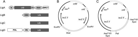
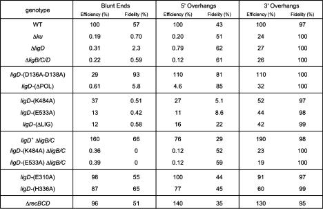
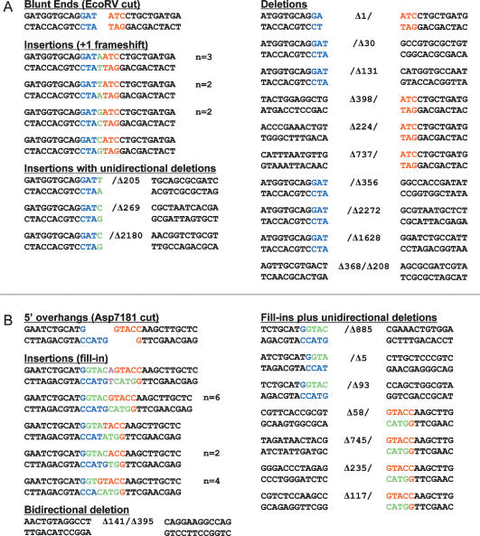
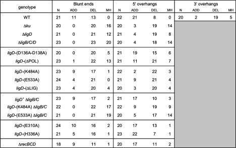
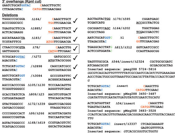
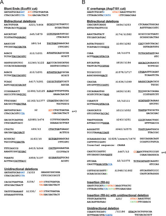
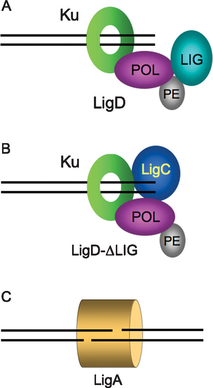
Mycobacteria can repair DNA double-strand breaks (DSBs) via a nonhomologous end-joining (NHEJ) system that includes a dedicated DNA ligase (LigD) and the DNA end-binding protein Ku. Here we exploit an improved plasmid-based NHEJ assay and a collection of Mycobacterium smegmatis strains bearing deletions or mutations in Ku or the DNA ligases to interrogate the contributions of LigD's three catalytic activities (polymerase, ligase, and 3' phosphoesterase) and structural domains (POL, LIG, and PE) to the efficiency and molecular outcomes of NHEJ in vivo. By analyzing in parallel the repair of blunt, 5' overhang, and 3' overhang DSBs, we discovered a novel end-joining pathway specific to breaks with 3' overhangs that is Ku- and LigD-independent and perfectly faithful. This 3' overhang NHEJ pathway is independent of ligases B and C; we surmise that it relies on NAD(+)-dependent LigA, the essential replicative ligase. We find that efficient repair of blunt and 5' overhang DSBs depends stringently on Ku and the LigD POL domain, but not on the LigD polymerase activity, which mainly serves to promote NHEJ infidelity. The lack of an effect of PE-inactivating LigD mutations on NHEJ outcomes, especially the balance between deletions and insertions at blunt or 5' overhang breaks, argues against LigD being the catalyst of deletion formation. Ligase-inactivating LigD mutations (or deletion of the LIG domain) have a modest impact on the efficiency of blunt and 5' overhang DSB repair, because the strand sealing activity can be provided in trans by one of the other resident ATP-dependent ligases (likely LigC). Reliance on the backup ligase is accompanied by a drastic loss of fidelity during blunt end and 5' overhang DSB repair. We conclude that the mechanisms of mycobacterial NHEJ are many and the outcomes depend on the initial structures of the DSBs and the available ensemble of end-processing and end-sealing components, which are not limited to Ku and LigD.


Figures

Figure 1.
Figure 1.
Mycobacterial ligases and new NHEJ reporter plasmids. (A) The mycobacterial LigA, LigB, LigC, and LigD polypeptides are depicted in cartoon form with the N termini on the left and the C termini on the right. Each enzyme has a core ligase catalytic domain (LIG) composed of a nucleotidyl transferase module and an OB-fold module. Flanking domains of known structure or function are shown. In LigA enzymes, the LIG domain is fused to an N-terminal domain (Ia) and three carboxyl domains: a tetracysteine Zn-finger, a helix–hairpin–helix (HhH) domain, and a BRCT domain. Domain Ia is unique to NAD+-dependent ligases and are required for the reaction of LigA with NAD+ to form the ligase-adenylate intermediate. LigB consists of a C-terminal LIG domain fused to an N-terminal DNA-binding domain (DBD). LigD and LigC are the NHEJ ligases of bacteria. LigC consists only of the catalytic core. LigD is a multifunctional repair protein that has N-terminal polymerase (POL) and central phosphoesterase (PE) catalytic modules fused to the C-terminal LIG domain. (B, C) Maps of the new NHEJ reporter plasmids, which contain a mycobacterial origin of replication (oriM), an E. coli origin of replication (oriE), a kanamycin resistance gene (kanR), and a lacZ gene interrupted by insertion of foreign DNA sequences between restriction sites for EcoRV (B) or the isoschizomers Asp7181/KpnI (C). To prepare the substrate for blunt-end NHEJ, the plasmid in B was digested with EcoRV and XbaI (which cuts uniquely within the foreign insert), and the linear EcoRV plasmid fragment was gel-purified. To prepare the substrates for 5′ overhang and 3′ overhang NHEJ, the plasmid in C was digested with either Asp7181 (5′ overhang) or KpnI (3′ overhang) plus PsiI (which cuts uniquely in the foreign insert), and the linear plasmid fragment was gel-purified.
Figure 2.
Figure 2.
NHEJ efficiency and fidelity in wild-type and mutant M. smegmatis strains. Genotypes are indicated in the left column. NHEJ efficiencies for repair of transfected linear plasmids with blunt, 5′ overhang, and 3′ overhang DSBs in the M. smegmatis mutants were normalized to the wild-type values (100%). NHEJ fidelity is the fraction (percentage) of kanR transformants that were lacZ+. The fidelity values for wild-type M. smegmatis for repair of blunt, 5′ overhang, and 3′ overhang ends were calculated from counts of 4128, 2259, and 2626 kanR colonies, respectively. The fidelity values in the Δku mutant for blunt, 5′ overhang, and 3′ overhang NHEJ were calculated from counts of 143, 79, and 2169 kanR colonies. The fidelity values in the ΔligD strain for blunt, 5′ overhang, and 3′ overhang NHEJ were calculated from counts of 523, 1539, and 4953 kanR colonies. The fidelity values in the ΔligB/C/D strain for blunt, 5′ overhang, and 3′ overhang NHEJ were calculated from counts of 169, 126, and 1613 kanR colonies.
Figure 3.
Figure 3.
Outcomes of unfaithful blunt and 5′ overhang DSB repair in wild-type M. smegmatis. The nucleotide sequences of NHEJ junctions from independent lacZ kanR colonies that had been transformed with EcoRV-cut (A) or Asp7181-cut (B) linear plasmid DNAs are shown. The two halves of the original restriction sites are shown in blue and red. Inserted nucleotides are colored green (or magenta in B when a nontemplated nucleotide is added after a 5′ overhang fill-in). Deletions are denoted by Δ next to the number of nucleotides resected from either end. The number of times (n) that the identical junction sequences were recovered from independent transformants is indicated at right, when applicable.
Figure 4.
Figure 4.
Summary of unfaithful NHEJ events. M. smegmatis genotypes are indicated in the left column. The total number (N) of unfaithfully repaired junctions that were sequenced for each strain and DSB end configuration is specified, along with the number of repair events that entailed nucleotide additions (ADD) or deletions (DEL) at the repair junctions. The incidence of ligation at microhomologies (defined as two or more positions of nucleotide identity flanking the junction) is reported for each data set.
Figure 5.
Figure 5.
Outcomes of unfaithful 3′ overhang DSB repair in wild-type M. smegmatis. The nucleotide sequences of NHEJ junctions from 20 independent lacZ kanR colonies that had been transformed with KpnI-cut linear plasmid DNA are shown. The two halves of the original KpnI restriction site are shown in blue and red. Deletions are denoted by Δ next to the number of nucleotides resected from either end. Inserted nucleotides are colored green. Microhomologies at the junctions are underlined and depicted as resulting from nucleolytic resection and exposure of complementary 3′ extensions, which can anneal to the 3′ single-stranded tail of one of the original KpnI-cut ends. Four of the junctions had sequences inserted between the sealed plasmid ends; three of these sequences correspond to distant sites in the pMSG288-based plasmid. These inserted sequences (and their numerical coordinates in the plasmid) are indicated below the junctions. The junction highlighted in the box exemplifies a likely instance of cross-break templated nucleotide addition to a 3′ single-stranded tail to fill-in a gap prior to strand sealing. Other likely instances of cross-break fill-in are indicated by check marks.
Figure 6.
Figure 6.
Outcomes of unfaithful blunt and 5′ overhang DSB repair in Δku cells. The nucleotide sequences of NHEJ junctions from independent lacZ kanR colonies that had been transformed with EcoRV-cut (A) or Asp7181-cut (B) linear plasmid DNAs are shown. The two halves of the original restriction sites are shown in blue and red. Deletions are denoted by Δ next to the number of nucleotides resected from either end. Inserted nucleotides are colored green. Microhomologies at the junctions are underlined and depicted as resulting from nucleolytic resection and exposure of complementary 5′ extensions. The number of times (n) that the identical junction sequences were recovered from independent transformants is indicated at right, when applicable.
Figure 7.
Figure 7.
Multiple NHEJ pathways in mycobacteria. Two alternative pathways of Ku-dependent blunt DSB repair are depicted in A and B. (A) In wild-type cells, the NHEJ apparatus is assembled at the DSB via contacts between the Ku homodimer (which encircles the DNA as a toroidal clamp) and the POL domain of LigD. The choice between faithful and unfaithful DSB repair reflects a kinetic balance between immediate sealing by the LIG domain (in the context of a synaptic complex with the other DSB end), nontemplated single nucleotide addition by the LigD POL domain, and end resection by an unknown mycobacterial nuclease. (B) Absent the LigD LIG domain (or when LigD ligase activity is suppressed), LigC provides backup sealing activity for blunt and 5′ overhang NHEJ. Because all blunt/5′overhang NHEJ requires the POL domain, we surmise that LigC interacts with the Ku–POL complex. Virtually all NHEJ events that proceed via this pathway are mutagenic. (C) M. smegmatis has a Ku-independent NHEJ pathway specific to sticky 3′ overhang DSBs that is nonmutagenic and, being active in ΔligB/C/D cells, apparently relies on LigA. It is not known if other accessory factors participate in this novel pathway.

Comment in

References

    1. Akey D., Martins A., Aniukwu J., Glickman M.S., Shuman S., Berger J.M. Crystal structure and nonhomologous end joining function of the ligase domain of Mycobacterium DNA ligase D. J. Biol. Chem. 2006;281:13412–13423. - PubMed
    1. Braunstein M., Brown A.M., Kurtz S., Jacobs W.R. Two nonredundant SecA homologues function in mycobacteria. J. Bacteriol. 2001;183:6979–6990. - PMC - PubMed
    1. Brissett N.C., Pitcher R.S., Juarez R., Picher A.J., Green A.J., Dafforn T.R., Fox G.C., Blanco L., Doherty A.J. Structure of a NHEJ polymerase-mediated synaptic complex. Science. 2007;318:456–459. - PubMed
    1. di d’Adda Fagagna F., Weller G.R., Doherty A.J., Jackson S.P. The Gam protein of bacteriophage Mu is an orthologue of eukaryotic Ku. EMBO Rep. 2003;4:47–52. - PMC - PubMed
    1. Della M., Palmbos P.L., Tseng H.M., Tonkin L.M., Daley J.M., Topper L.M., Pitcher R.S., Tomkinson A.E., Wilson T.E., Doherty A.J. Mycobacterial Ku and ligase proteins constitute a two-component NHEJ repair machine. Science. 2004;306:683–685. - PubMed

Publication types

MeSH terms