. 2008 Feb 15;68(4):981-8.
doi: 10.1158/0008-5472.CAN-06-0249.

Growth suppression of lung cancer cells by targeting cyclic AMP response element-binding protein

Affiliations

Growth suppression of lung cancer cells by targeting cyclic AMP response element-binding protein

Sita Aggarwal et al. Cancer Res. .

Abstract

Genes regulated by cyclic AMP-response element-binding protein (CREB) have been reported to suppress apoptosis, induce cell proliferation, and mediate inflammation and tumor metastasis. However, it is not clear whether CREB is critically involved in lung carcinogenesis. We found that non-small cell lung cancer (NSCLC) cell lines exhibited elevated constitutive activity in CREB, in its immediate upstream kinases (ribosomal s6 kinase and extracellular signal kinase), and in the CREB-regulated cell survival proteins Bcl-2 and Bcl-xL. We hypothesized that constitutively active CREB is important to lung cancer cell growth and survival and therefore could be a potential therapeutic target for NSCLC. Ectopic expression of dominant repressor CREB and transfection with small interfering RNA against CREB suppressed the growth and survival of NSCLC cells and induced apoptotic cell death. Furthermore, treating H1734 NSCLC cells with an inhibitor of the CREB signaling pathway Ro-31-8220 inhibited CREB activation by blocking the activity of extracellular signal kinase and ribosomal s6 kinase, arrested the cell cycle at the G(2)-M phase, and subsequently induced apoptosis with the suppression of Bcl-2 and Bcl-xL expression. Ro-31-8220 suppressed both the anchorage-dependent and independent growth of NSCLC cells, but its cytotoxic effect was much less prominent in normal bronchial epithelial cells. Our results indicate that active CREB plays an important role in NSCLC cell growth and survival. Thus, agents that suppress CREB activation could have potential therapeutic value for NSCLC treatment.


Figures
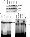
Figure 1
Figure 1
CREB was overexpressed and constitutively activated in NSCLC cells. A, Whole cell lysates were made from NHTBE, H1734, H226, H292, A549 cells and tested for constitutive levels of pCREB and total CREB by western blot analysis. The same blot was stripped and re-probed with an anti-β-actin antibody to show equal loading of samples (lower panel). B, Nuclear extracts from NHTBE, H1734, A549, H226, and H292 cells were prepared as described in Methods and then assayed for CREB activation by EMSA with a radio-labeled CRE consensus oligonucleotide probe. C, To verify the specificity of EMSA analysis, nuclear extracts from H1734 cells were incubated with CRE probe in the presence of anti-pCREB or anti-CREB antibodies or 100 fold unlabeled (cold) CRE oligonucleotide or mutant CRE oligonucleotide or non specific Immunoglobin G (IgG), and then the band pattern change was examine in EMSA. *: Shifted band. The representative gel images from three independent experiments are shown.
Figure 2
Figure 2
Ro-31-8220 inhibited constitutively active CREB in NSCLC cells. A. H1734 cells were treated with 20 μM Ro-31-8220 for the designated times, or with various concentrations of Ro-31-8220 for 4 h. The whole cell extracts were analyzed with western blot using anti p-CREB. In each case, the same blot was stripped and re-probed with the anti-CREB antibody (lower panel in each figure). B. The nuclear extract from H1734 cells received the above treatments were subjected to EMSA for CRE-binding ability. C. Ro-31-8220 induced the disappearance of nuclear pCREB in NSCLC cells. H1734 cells were incubated with or without 20 μM Ro-31-8220 for 4 h and then analyzed for pCREB with immunocytochemistry. The green stain indicates the localization of pCREB, and the blue stain indicates the localization of the nucleus. The images shown are representatives of three independent experiments.
Figure 3
Figure 3
Upstream and downstream components of CREB signaling are constitutively over-activated in NSCLC cells and their activation is suppressed by Ro-31-8220. A. Whole cell lysates from NSCLC cell lines (H1734, H226, H292, and A549) and NHTBE cells were analyzed with western blot for the levels of phospho-Rsk and phospho-Erk1/2, and the expression of cell survival gene, Bcl-2 and Bcl-xL. B. and C. H1734 cells were treated with 20 μM Ro-31-8220 for the indicated times or with various concentrations of Ro-31-8220 for 4 h, and the whole cell lysate was assessed for the levels of signaling components mentioned above. Equal sample loading is indicated by the band intensity of the inactive form of each protein or β-actin (lower panel in each figure).
Figure 4
Figure 4
Ro-31-8220 suppresses cell growth and colony formation in NSCLC cells. A, Cells (H1734, H226, A549 and H292) were plated in 96-well plates and incubated in triplicate with the indicated dose of Ro-31-8220 for different days. Cell proliferation was assessed using MTT assays. The results are shown as the means (± standard error) of three independent experiments. B, The effect of Ro-31-8220 (10 μM) on cell viability was compared between NHTBE and NSCLC (H1734 and H226) cells using MTT assay as mentioned above. The results are shown as the means (± standard error) of three independent experiments. C. H1734 and H226 cells were seeded in 0.35% agar and treated with different concentrations of Ro-31-8220. Colonies formed after 28-day incubation at 37°C in 5% CO2 were counted under light microscopy. Colonies greater than 60 μm were scored. Result is mean (± standard error) of three independent experiments. **: p<0.01.
Figure 5
Figure 5
Ro-31-8220 leads to G2/M cell cycle arrest followed by apoptosis in NSCLC cells. A, Serum-starvation synchronized H1734 cells and NHTBE cells were incubated in the absence or presence of 10 μM Ro-31-8220 for the indicated times. Then, cells were washed, fixed, stained with propidium iodide, and analyzed for DNA content by flow cytometry. Data (means ± standard error) shown are percentages of cells in each phase of cell cycle from three experiments. *: p<0.05; **: p<0.01 (one-tailed, t-test). B, H1734 cells were incubated with or without 20 μM Ro-31-8220 for the indicated times in medium containing 5% of serum. Whole cell lysates were subjected to western blot analysis using anti-PARP, anti-caspase-9 antibodies. C, Suppression of Ro-31-8220 induced caspase-3 cleavage by caspase-3 inhibitor. H1734 cells were pre-incubated with and without caspase inhibitor 5 μM Ac-DEVD-CHO for 2 h and then treated with 20 μM Ro-31-8220 for 24 h. Then, cell extracts were prepared and analyzed for caspase-3 cleavage by Western blot analysis using an anti-caspase-3 antibody. DMSO was used as a vehicle control. D, The caspase-3 inhibitor protects cells from Ro-31-8220 induced cytotoxicity. H1734 cells were incubated with different concentrations of caspase inhibitor Ac-DEVD-CHO as indicated for 2 h and then treated with 20 μM Ro-31-8220. After 24 h, cell viability was determined by the MTT. The results are shown as the means (± standard error) percentage viability from triplicate cultures of three independent experiments. **: p<0.01.
Figure 6
Figure 6
Suppression of CREB inhibits cell proliferation and induces apoptosis. A, H1734 cells were transfected with CREBwt, KCREB, and CREB133 plasmid, and then assayed for cell proliferation by the MTT assays. The results are shown as the means (± standard error) of three independent experiments. B, H1734 cells were transfected with siCREB and non-specific control pool siRNA (NS-siRNA), then assayed for cell proliferation by the MTT assays. The results are shown as the means (± standard error) of three independent experiments. C, H1734 cells were transiently transfected with siCREB, siRNA-NS, CREBwt, KCREB, and CREB133 plasmid. Three days after transfection, equal amounts of whole-cell lysates were prepared and subjected to western blot analysis using the indicated antibodies. D, TUNEL analysis was performed on H1734 cells 48 h after transfection with the indicated plasmids or treatment with Ro-31-8220 (5 μM). Nuclei appear blue (DAPI stain), TUNEL-positive nuclei stain green. Magnification: 200x. The results are shown as the means of three independent experiments.

References

    1. Lynch TJ, Bell DW, Sordella R, et al. Activating mutations in the epidermal growth factor receptor underlying responsiveness of non-small-cell lung cancer to gefitinib. N Engl J Med. 2004;350:2129–39. - PubMed
    1. Paez JG, Janne PA, Lee JC, et al. EGFR mutations in lung cancer: correlation with clinical response to gefitinib therapy. Science. 2004;304:1497–500. - PubMed
    1. Sequist LV, Bell DW, Lynch TJ, Haber DA. Molecular predictors of response to epidermal growth factor receptor antagonists in non-small-cell lung cancer. J Clin Oncol. 2007;25:587–95. - PubMed
    1. Conkright MD, Montminy M. CREB: the unindicted cancer co-conspirator. Trends Cell Biol. 2005;15:457–9. - PubMed
    1. Riccio A, Ahn S, Davenport CM, Blendy JA, Ginty DD. Mediation by a CREB family transcription factor of NGF-dependent survival of sympathetic neurons. Science (New York, NY. 1999;286:2358–61. - PubMed

Publication types

MeSH terms