. 2008 Apr 15;111(8):4118-25.
doi: 10.1182/blood-2007-12-127928. Epub 2008 Feb 15.

Vascular dermatan sulfate regulates the antithrombotic activity of heparin cofactor II

Affiliations

Vascular dermatan sulfate regulates the antithrombotic activity of heparin cofactor II

Li He et al. Blood. .

Abstract

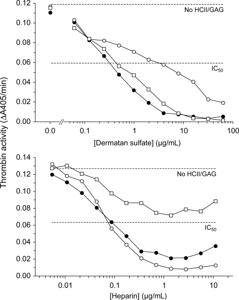
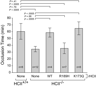
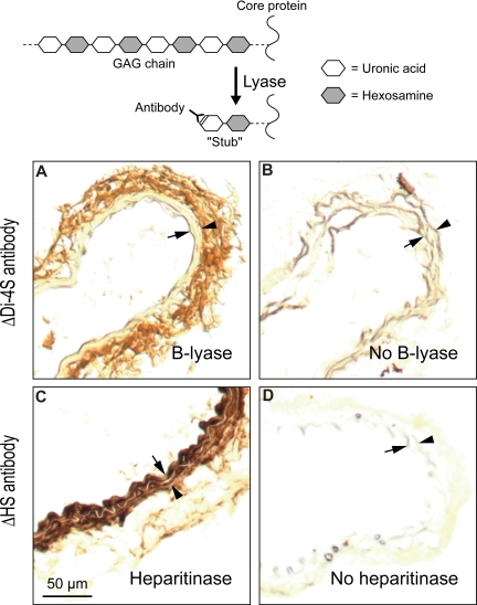
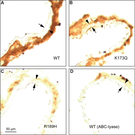
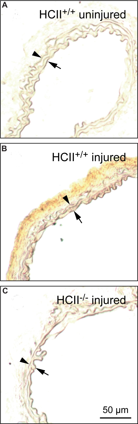
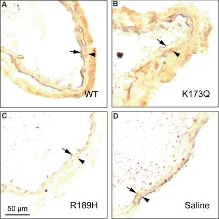
Heparin cofactor II (HCII)-deficient mice form occlusive thrombi more rapidly than do wild-type mice following injury to the carotid arterial endothelium. Dermatan sulfate (DS) and heparan sulfate (HS) increase the rate of inhibition of thrombin by HCII in vitro, but it is unknown whether vascular glycosaminoglycans play a role in the antithrombotic effect of HCII in vivo. In this study, we found that intravenous injection of either wild-type recombinant HCII or a variant with low affinity for HS (K173H) corrected the abnormally short thrombosis time of HCII-deficient mice, while a variant with low affinity for DS (R189H) had no effect. When HCII was incubated with frozen sections of the mouse carotid artery, it bound specifically to DS in the adventitia. HCII was undetectable in the wall of the uninjured carotid artery, but it became concentrated in the adventitia following endothelial injury. These results support the hypothesis that HCII interacts with DS in the vessel wall after disruption of the endothelium and that this interaction regulates thrombus formation in vivo.


Figures

Figure 1
Figure 1
Inhibition of murine thrombin by rHCII variants. Murine thrombin (14 nM) was incubated for 60 seconds with 185 nM human rHCII (WT, ●; R189H, ○; K173Q, □) and either DS or heparin at the final concentrations shown. Residual thrombin activity was then assayed with a chromogenic substrate (ΔA405/min). No HCII/GAG indicates thrombin activity in the absence of HCII and glycosaminoglycan; IC50, 50% of initial thrombin activity.
Figure 2
Figure 2
Thrombotic occlusion times after photochemical injury of the carotid artery. HCII+/+ or HCII−/− mice were injected intravenously with saline (none) or purified rHCII (WT, R189H, or K173Q) 15 minutes before injection of rose bengal dye. The dose of rHCII was calculated to achieve a plasma concentration of 0.125 μM. The bars indicate the means plus or minus SD.
Figure 3
Figure 3
Clearance of rHCII from the mouse circulation. HCII−/− mice were injected intravenously with rHCIIWT (●, —) or rHCIIR189H (○, ----) as in Figure 2. Blood samples were collected 5 minutes, 1 hour, and 2 hours after injection, and the plasma HCII antigen was determined in duplicate by ELISA. HCII antigen values were normalized to the 5-minute point (100%). The half-life was calculated from the slope of the line fit to log(HCII antigen) versus time and was approximately 0.9 hour for each protein.
Figure 4
Figure 4
Localization of DS and HS in uninjured carotid arteries of wild-type mice. Frozen sections were treated with chondroitin B-lyase (A), Flavobacterium heparitinase (C), or buffer alone (B,D) and then incubated with monoclonal antibodies ΔDi-4S (A,B) or ΔHS (C,D). Bound monoclonal antibodies were detected with a peroxidase-conjugated secondary antibody. Arrow indicates internal elastic lamina; arrowhead, external elastic lamina. DS was present primarily in the adventitia (A) and HS in the intima/media (C).
Figure 5
Figure 5
Binding of HCII to carotid arterial sections in vitro. Frozen sections were incubated for 1 hour at room temperature with 20 μg/mL rHCIIWT (A), rHCIIK173Q (B), or rHCIIR189H (C) in PBS. After 2 rinses with buffer, the sections were stained with a polyclonal goat anti-HCII IgG. The section in panel D was treated with chondroitin ABC-lyase to degrade DS prior to incubation with rHCIIWT. Arrow indicates internal elastic lamina; arrowhead, external elastic lamina. rHCIIWT and rHCIIK173Q bound strongly to sites in the adventitia.
Figure 6
Figure 6
Distribution of endogenous HCII before and after injury. Frozen sections of carotid arteries harvested from HCII+/+ mice before (A) or 30 minutes after (B) the onset of injury were stained with a polyclonal goat anti–HCII IgG. No HCII antigen was detected in the injured carotid artery of a control (HCII−/−) mouse (C). Arrow indicates internal elastic lamina; arrowhead, external elastic lamina.
Figure 7
Figure 7
Distribution of rHCII variants after arterial injury. HCII−/− mice were injected with rHCIIWT (A), rHCIIK173Q (B), rHCIIR189H (C), or saline (D) and then subjected to photochemical injury as described in the legend to Figure 2. Frozen sections of arteries harvested 30 minutes after the onset of injury were stained with a polyclonal goat anti–HCII IgG. Arrow indicates internal elastic lamina; arrowhead, external elastic lamina.

Similar articles

Cited by

References

    1. Mann KG. Thrombin formation. Chest. 2003;124:4S–10S. - PubMed
    1. Coughlin SR. Protease-activated receptors in hemostasis, thrombosis and vascular biology. J Thromb Haemost. 2005;3:1800–1814. - PubMed
    1. Bock S. Antithrombin III and heparin cofactor II. In: Colman R, Marder V, Clowes A, George J, Goldhaber S, editors. Hemostasis and Thrombosis: Basic Principles and Clinical Practice. 5th ed. Philadelphia, PA: Lippincott Williams & Wilkins; 2006. pp. 235–248.
    1. Tollefsen D, Zhang L. Heparin and vascular proteoglycans. In: Colman R, Marder V, Clowes A, George J, Goldhaber S, editors. Hemostasis and Thrombosis: Basic Principles and Clinical Practice. 5th ed. Philadelphia, PA: Lippincott Williams & Wilkins; 2006. pp. 271–283.
    1. Tollefsen DM. Antithrombin deficiency. In: Scriver C, Beaudet A, Sly W, et al., editors. The Metabolic and Molecular Bases of Inherited Disease. 8th ed. New York, NY: McGraw-Hill; 2001. pp. 4455–4471.

Publication types

MeSH terms