. 2008 Apr;36(7):2208-18.
doi: 10.1093/nar/gkn060. Epub 2008 Feb 16.

Nucleosome formation with the testis-specific histone H3 variant, H3t, by human nucleosome assembly proteins in vitro

Affiliations

Nucleosome formation with the testis-specific histone H3 variant, H3t, by human nucleosome assembly proteins in vitro

Hiroaki Tachiwana et al. Nucleic Acids Res. 2008 Apr.

Abstract

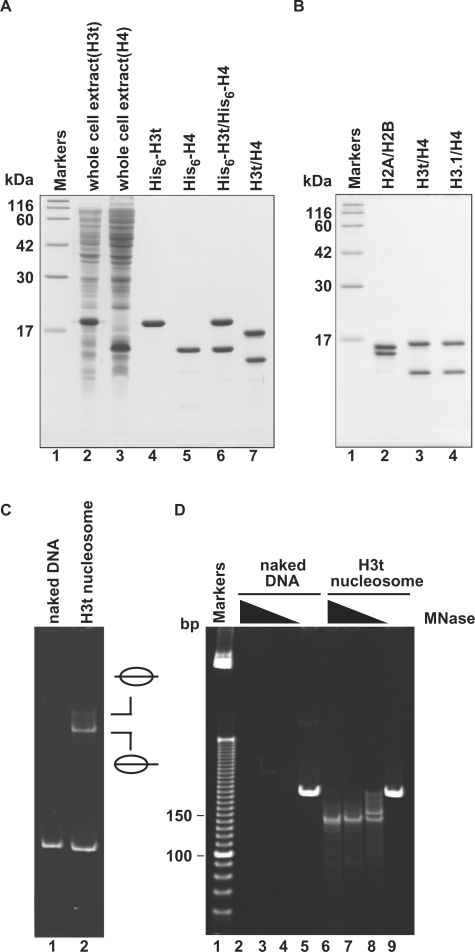
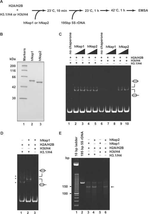
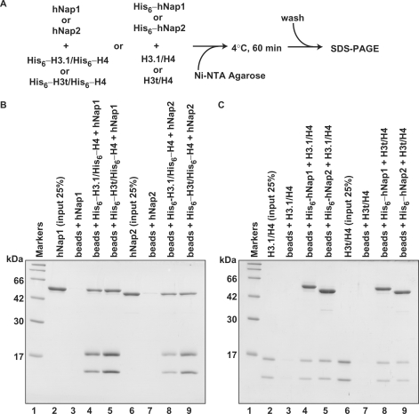
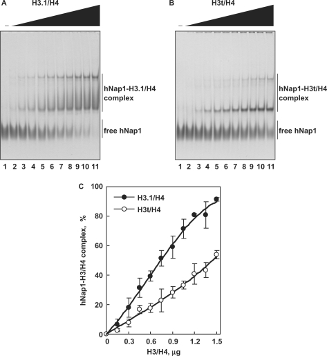
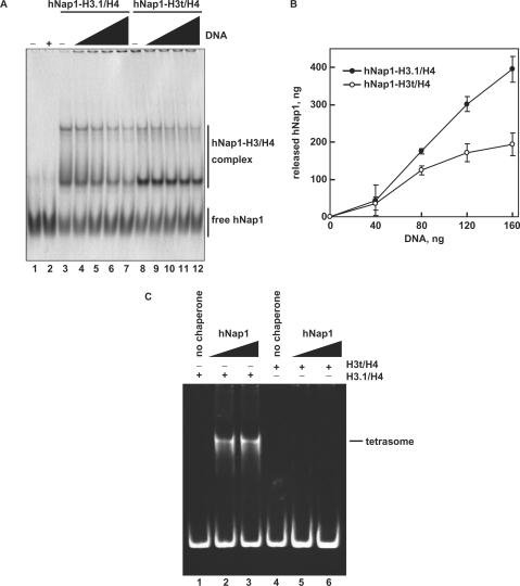
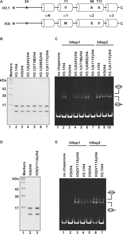
Five non-allelic histone H3 variants, H3.1, H3.2, H3.3, H3t and CENP-A, have been identified in mammals. H3t is robustly expressed in the testis, and thus was assigned as the testis-specific H3 variant. However, recent proteomics and tissue-specific RT-PCR experiments revealed a small amount of H3t expression in somatic cells. In the present study, we purified human H3t as a recombinant protein, and showed that H3t/H4 forms nucleosomes with H2A/H2B by the salt-dialysis method, like the conventional H3.1/H4. We found that H3t/H4 is not efficiently incorporated into the nucleosome by human Nap1 (hNap1), due to its defective H3t/H4 deposition on DNA. In contrast, human Nap2 (hNap2), a paralog of hNap1, promotes nucleosome assembly with H3t/H4. Mutational analyses revealed that the Ala111 residue, which is conserved among H3.1, H3.2 and H3.3, but not in H3t, is the essential residue for the hNap1-mediated nucleosome assembly. These results suggest that H3t may be incorporated into chromatin by a specific chaperone-mediated pathway.


Figures

Figure 1.
Figure 1.
Preparation and nucleosome formation ability of H3t/H4. (A) SDS–PAGE analysis of proteins from each preparation step for H3t/H4. Lanes 2 and 3: the whole cell lysates of the E. coli cells expressing His6-tagged H3t and His6-tagged H4, respectively. Lanes 4 and 5: the peak Ni-NTA agarose fractions of His6-tagged H3t and His6-tagged H4, respectively. Lanes 6 and 7: His6-tagged H3t/H4 before and after the removal of the hexahistidine tag, respectively. The positions of the size markers (lane1) are indicated on the left. (B) SDS–PAGE analysis of purified histones used in this study. Lane 1: size markers. Lanes 2–4: histones; H2A/H2B (lane 2), H3t/H4 (lane 3) and H3.1/H4 (lane 4). (C) Nucleosome reconstitution analyzed by non-denaturing 6% PAGE. Lane 1: naked DNA. Lane 2: nucleosomes reconstituted with H2A/H2B and H3t/H4 by the salt-dialysis method. Nucleosomes with different translational positions are indicated. (D) MNase assay analyzed by non-denaturing 10% PAGE. Naked DNA (lanes 2–5) and H3t-containing nucleosomes reconstituted by the salt-dialysis method (lanes 6–9) were treated with MNase, and the resulting DNA fragments were analyzed. Lane 1: the molecular mass markers (10 bp DNA ladder). Amounts of MNase (unit/μl) were 0.18 (lanes 2 and 6), 0.13 (lanes 3 and 7), 0.08 (lanes 4 and 8) and 0 (lanes 5 and 9). In H3t-containing nucleosomes, 146 bp DNA, which is tightly wrapped around the nucleosomes and resistant to MNase, is detected (lanes 6–8).
Figure 2.
Figure 2.
Nucleosome assembly with H2A/H2B and H3t/H4 or H3.1/H4 by hNap1 and hNap2. (A) Schematic representation of the nucleosome-reconstitution assay by hNap1 or hNap2. (B) SDS–PAGE analysis of purified hNap1 (lane 2) and hNap2 (lane 3) lacking the His6 tag. (C) Nucleosome-reconstitution experiments with the 42°C incubation, analyzed by non-denaturing 6% PAGE. A total of 195 bp 5S DNA was incubated with histones without (lanes 1 and 6) or in combination with hNap1 (lanes 2–3 and 7–8), or hNap2 (lanes 4–5 and 9–10). To assemble nucleosomes containing the histone octamer, H2A/H2B was used with H3.1/H4 (lanes 1–5) or H3t/H4 (lanes 6–10). (D) Nucleosome-reconstitution experiments without the 42°C incubation, analyzed by non-denaturing 6% PAGE. Reactions were performed as in (C). Lane 1 indicates the experiment with H3.1/H4/H2A/H2B in the absence of hNap1. Lanes 2 and 3 indicate the experiments with H3.1/H4/H2A/H2B and H3t/H4/H2A/H2B, respectively, in the presence of hNap1. Bands corresponding to non-specific DNA binding of histones are indicated by asterisks. (E) MNase assay analyzed by non-denaturing 10% PAGE. The samples incubated with the indicated combinations were treated with MNase, and the resulting DNA fragments were analyzed. A total of 146 bp DNA fragments in nucleosomes (arrow) are found in the combinations of H3.1/H4 with hNap1 (lane 3) or hNap2 (lane 4), and H3t/H4 with hNap2 (lane 6), but not H3t/H4 with hNap1 (lane 5).
Figure 3.
Figure 3.
The Ni-NTA-agarose pull-down assay. (A) A schematic diagram of the assay. (B) Pull-down assay using His6-tagged histones. hNap1 (lanes 3–5) or hNap2 (lanes 7–9) was mixed with His6-tagged H3.1/H4 (lanes 4 and 8) or His6-tagged H3t/H4 (lanes 5 and 9), and the proteins bound to the Ni-NTA agarose beads were analyzed by 15% SDS–PAGE with CBB staining. Lanes 2 and 6: the inputs (25%) of hNap1 and hNap2, respectively. (C) Pull-down assay using His6-tagged hNap1 and hNap2. H3.1/H4 (lanes 3–5) or H3t/H4 (lanes 7–9) was mixed with His6-tagged hNap1 (lanes 4 and 8) or His6-tagged hNap2 (lanes 5 and 9), and the proteins bound to the Ni-NTA agarose beads were analyzed by 15% SDS–PAGE with CBB staining. Lanes 2 and 6: the inputs (25%) of H3.1/H4 and H3t/H4, respectively.
Figure 4.
Figure 4.
The gel mobility shift assay. (A) hNap1-H3.1/H4 complex formation. hNap1 (2.3 μg) was incubated with different amounts of H3.1/H4, and the hNAP1-H3.1/H4 complex was detected by non-denaturing 5% PAGE with CBB staining. The amounts of H3.1/H4 (μg) used in this study were: 0 (lane 1), 0.15 (lane 2), 0.3 (lane 3), 0.45 (lane 4), 0.6 (lane 5), 0.75 (lane 6), 0.9 (lane 7), 1.05 (lane 8), 1.2 (lane 9), 1.35 (lane 10) and 1.5 (lane 11). (B) hNap1-H3t/H4 complex formation. hNap1 (2.3 μg) was incubated with different amounts of H3t/H4, and the hNap1-H3t/H4 complex formation was analyzed as in (A). (C) Graphic representation of the hNap1-H3/H4complex formation. The relative amounts of hNap in the complex with H3.1/H4 (closed circles) and H3t/H4 (open circles) were plotted as the averages of three independent experiments, as in (A) and (B), with the SD values.
Figure 5.
Figure 5.
The H3/H4 deposition assay. (A) The deposition of H3.1/H4 or H3t/H4 from the hNap1-H3.1/H4 or hNap1-H3t/H4 complex onto DNA, analyzed by non-denaturing 5% PAGE with CBB staining. hNap1 (2.3 μg) was incubated without (lanes 1–2) or with H3.1/H4 (0.75 μg; lanes 3–7) or H3t/H4 (1.2 μg; lanes 8–12) to form the complex; about half of the hNap1 remained free under these conditions (lanes 3 and 8). After the incubation with supercoiled DNA, the samples were analyzed. The amounts of competitor DNA (ng) were 0 (lanes 3 and 8), 40 (lanes 4 and 9), 80 (lanes 5 and 10), 120 (lanes 6 and 11) and 160 (lanes 2, 7 and 12). (B) Graphic representation of hNap1 release. The amounts of hNap1 released from hNap1-H3.1/H4 (open circles) or hNap1-H3t/H4 complex (closed circles) are plotted as the averages of three independent experiments, performed as in (A), with the SD values. (C) Tetrasome-reconstitution with H3.1/H4 or H3t/H4 by hNap1. A total of 195 bp 5S DNA was incubated with hNap1 in combination with H3.1/H4 (8 ng/μl; lanes 1–3) or H3t/H4 (8 ng/μl; 4–6) (in the absence of H2A/H2B). The tetrasome formation was analyzed by non-denaturing 6% PAGE. The amounts of hNap1 (ng/μl) were 0 (lane 1), 45 (lane 2) and 182 (lane 3). The amounts of hNap2 (ng/μl) were 0 (lane 4), 43 (lane 5) and 171 (lane 6).
Figure 6.
Figure 6.
Mutational analyses of H3.1 and H3t. (A) Sequence comparison between human H3.1 and H3t. The different amino acids between H3.1 and H3t are indicated by capital letters. Cylinders indicate α-helices found in the crystal structure of the human nucleosome core particle (39). (B) SDS–PAGE analysis of the H3.1 mutants complexed with H4. Lane 1: molecular mass markers. Lanes 2 and 3: purified H3.1/H4 and H3t/H4, respectively. Lanes 4–7: H3.1 mutants complexed with H4; lane 4: H3.1(A24V), lane 5: H3.1(V71M), lane 6: H3.1(A98S) and lane 7: H3.1(A111V). (C) Nucleosome-reconstitution with the H3.1 mutants by hNAP1 or hNAP2, analyzed by non-denaturing 6% PAGE. A total of 195 bp 5S DNA was incubated with hNap1 (lanes 2–7) or hNap2 (lanes 8–10) in combination with H3.1/H4 (lanes 2 and 8), H3t/H4 (lanes 3 and 9) and H3.1 mutants/H4 (lanes 4–7 and 10); all reactions were performed in the presence of H2A/H2B. (D) SDS–PAGE analysis of the H3t(V111A) mutant complexed with H4. (E) Nucleosome-reconstitution with H3t(V111A)/H4 by hNap1 or hNap2, analyzed by non-denaturing 6% PAGE. A total of 195 bp 5S DNA was incubated with hNap1 (lanes 2–4) or hNap2 (lanes 5–7) in combination with H3t/H4 (lanes 2 and 5), H3t(V111A)/H4 (lanes 3 and 6) and H3.1/H4 (lanes 4 and 7); all reactions were performed in the presence of H2A/H2B.

References

    1. van Holde KE. Chromatin. New York: Springer; 1989.
    1. Wolffe AP. Chromatin: Structure & Function. London: Academic Press; 1998.
    1. Richmond TJ, Finch JT, Rushron B, Rhodes D, Klug A. Structure of the nucleosome core particle at 7 Å resolution. Nature. 1984;311:532–537. - PubMed
    1. Luger K, Mader AW, Richmond RK, Sargent DF, Richmond TJ. Crystal structure of the nucleosome core particle at 2.8 Å resolution. Nature. 1997;389:251–260. - PubMed
    1. Arents G, Moudrianakis EN. The histone fold: a ubiquitous architectural motif utilized in DNA compaction and protein dimerization. Proc. Natl Acad. Sci. USA. 1995;92:11170–11174. - PMC - PubMed

Publication types