. 2008 Feb 8;4(2):e29.
doi: 10.1371/journal.ppat.0040029.

A mouse model for Chikungunya: young age and inefficient type-I interferon signaling are risk factors for severe disease

Affiliations

A mouse model for Chikungunya: young age and inefficient type-I interferon signaling are risk factors for severe disease

Thérèse Couderc et al. PLoS Pathog. .

Abstract

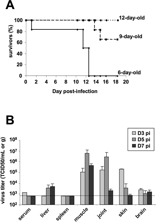
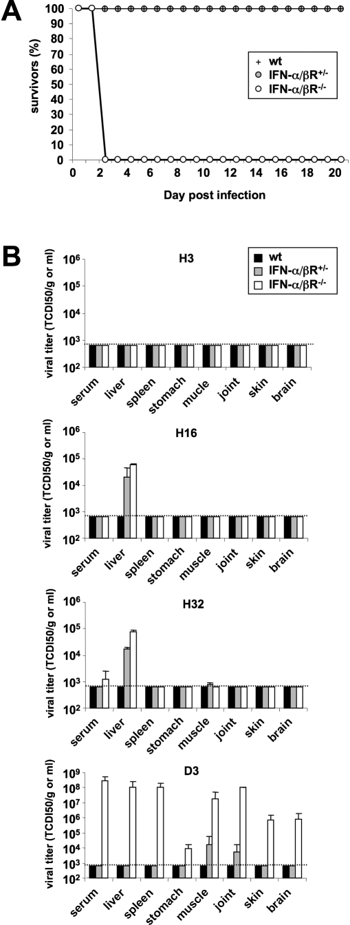
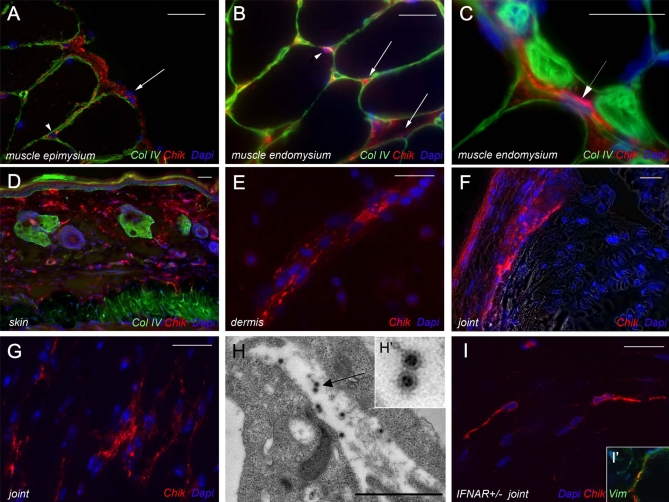
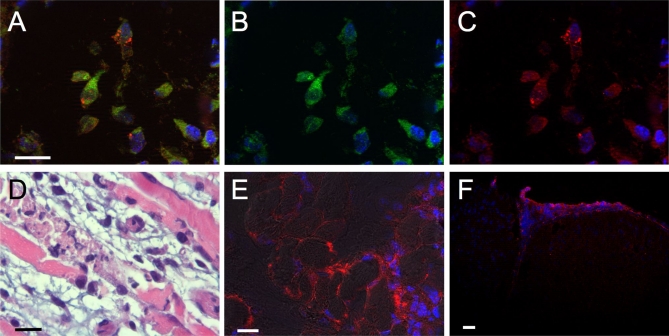
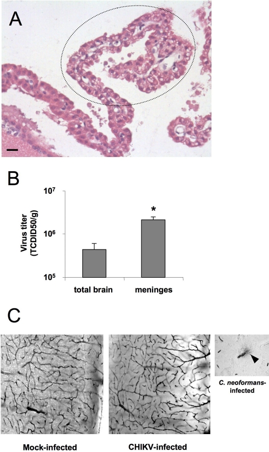
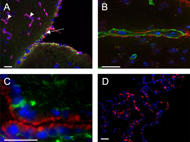
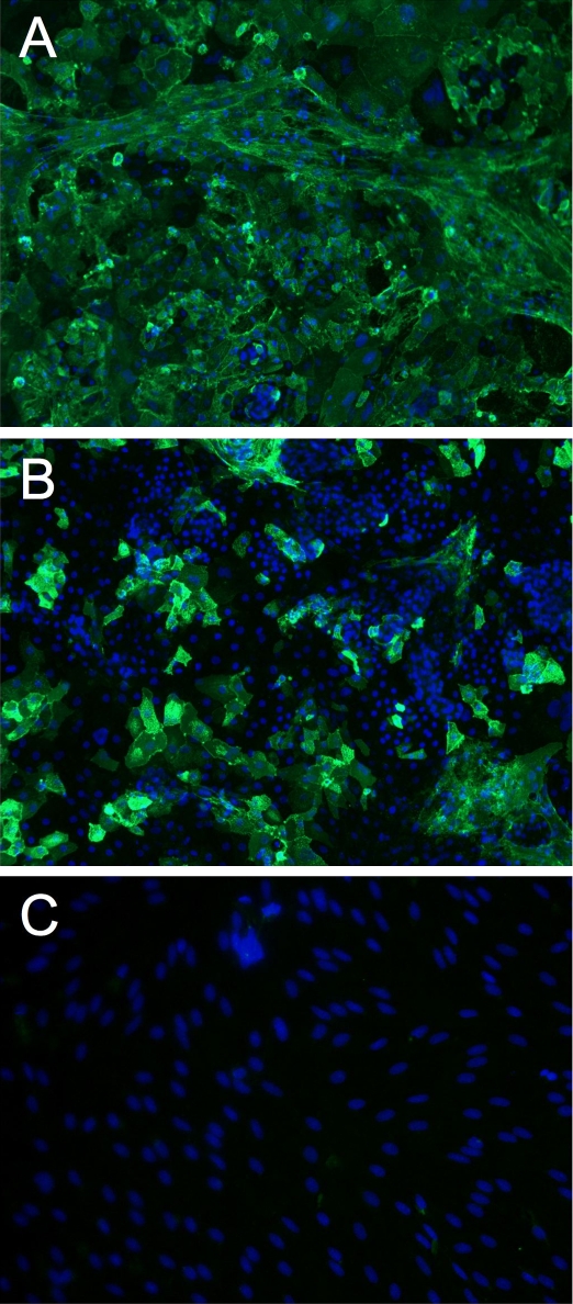
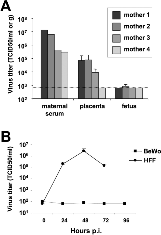
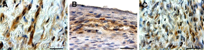
Chikungunya virus (CHIKV) is a re-emerging arbovirus responsible for a massive outbreak currently afflicting the Indian Ocean region and India. Infection from CHIKV typically induces a mild disease in humans, characterized by fever, myalgia, arthralgia, and rash. Cases of severe CHIKV infection involving the central nervous system (CNS) have recently been described in neonates as well as in adults with underlying conditions. The pathophysiology of CHIKV infection and the basis for disease severity are unknown. To address these critical issues, we have developed an animal model of CHIKV infection. We show here that whereas wild type (WT) adult mice are resistant to CHIKV infection, WT mouse neonates are susceptible and neonatal disease severity is age-dependent. Adult mice with a partially (IFN-alpha/betaR(+/-)) or totally (IFN-alpha/betaR(-/-)) abrogated type-I IFN pathway develop a mild or severe infection, respectively. In mice with a mild infection, after a burst of viral replication in the liver, CHIKV primarily targets muscle, joint, and skin fibroblasts, a cell and tissue tropism similar to that observed in biopsy samples of CHIKV-infected humans. In case of severe infections, CHIKV also disseminates to other tissues including the CNS, where it specifically targets the choroid plexuses and the leptomeninges. Together, these data indicate that CHIKV-associated symptoms match viral tissue and cell tropisms, and demonstrate that the fibroblast is a predominant target cell of CHIKV. These data also identify the neonatal phase and inefficient type-I IFN signaling as risk factors for severe CHIKV-associated disease. The development of a permissive small animal model will expedite the testing of future vaccines and therapeutic candidates.


Conflict of interest statement

Competing interests. The authors have declared that no competing interests exist.

Figures

Figure 1
Figure 1. CHIKV Infection in Mouse Neonates
(A) Survival of mouse neonates according to their age. Mouse neonates were inoculated with 106 PFU of CHIKV via the ID route and observed for lethality (n = 6 per group). (B) Viral titers in tissues of 9-day-old neonates. Mice were inoculated with 106 PFU of CHIKV via the ID route and sacrificed at the indicated time points. The amount of infectious virus in serum and tissues were quantified by TCID50. Each data point represents the arithmetic mean ± SD for at least four mice. The broken line indicates the detection threshold.
Figure 2
Figure 2. CHIKV Infection in Mice According to the Integrity of the IFNα/β Pathway
(A) Survival of mice inoculated with 106 PFU of CHIKV via the ID route (n = 6 for each category). (B) Viral titers in IFN-α/βR−/−, IFN-α/βR+/−, and WT adult mice infected via the ID route with 20, 106, and 106 PFU, respectively. At the indicated time points, mice were sacrificed and the amount of infectious virus present in serum and tissues was quantified by TCID50. Each data point represents the arithmetic mean ± SD for at least four mice. A broken line indicates the detection threshold.
Figure 3
Figure 3. CHIKV Cell and Tissue Tropisms in Mice
IFN-α/βR−/− mice ID inoculated with 20 PFU and IFN-α/βR+/− ID inoculated with 106 PFU route were sacrificed at D3 pi. Multiple immunostaining were performed on tissue cryosections from IFN-α/βR−/− mice (A to G) and from IFN-α/βR+/− mouse (I), and transmission electron microscopy on joint from IFN-α/βR−/− mice (H). Nuclei stained by Hoechst appear in blue, CHIKV in red (A to G and I). Basal lamina (collagen IV) (A to D) and mesenchymal cells (vimentin) (I′) appear in green. In all these pictures bar is 10 μm. Muscle fibroblasts, identified notably as cells not surrounded by a basal lamina, display a strong immunostaining for CHIKV in the epimysium (arrow) and endomysium (arrowhead) of skeletal muscle (A). Fibroblasts (arrows) and very few satellite cells (arrowhead) easily recognizable as a single mononucleated cell located beneath the muscle fiber basal lamina are labeled for CHIKV antigens in endomysium of skeletal muscle (B and C). Fibroblasts were also immunostained for CHIKV in deep dermis (D and E) and in joint capsule (F and G). (H) Transmission electron microcopy view of viral particles (arrow in H and H′) in contact and within the cytoplasm of a cell in connective tissue, identified as a fibroblast because of the absence of surrounding basal lamina and the adjacent type I collagen fibers. Infected cells positive for CHIKV in skeletal muscle of IFN-α/βR+/− mouse (I), identified as fibroblasts since they were not surrounded by basal lamina and expressed vimentin (I′).
Figure 4
Figure 4. CHIKV Cell and Tissue Tropisms in Mouse Neonates
Nine-day old IFN-α/βR+/+ mice were infected with 106 PFU of CHIKV by ID route. Overlay image (A) of infected muscle connective tissue immunolabeled for vimentin (B) and CHIKV (C). Hematoxylin and eosin staining (D) of longitudinal section of skeletal muscle shows a severe necrotizing myositis with numerous infiltrates and necrosis of the muscle fibers. Immunostaining of CHIKV antigens (red) and nuclei (blue) on muscle (E) and brain (F) sections. Note that the endomysium displays a strong CHIKV immunolabeling, as well as leptomeningeal cells. Bar is 10μm.
Figure 5
Figure 5. CHIKV Targets the Choroid Plexus and Meningeal Tissue but Does Not Affect the BBB Permeability in IFN-α/βR−/− Mice
Mice were infected with 20 PFU of CHIKV via the ID route and sacrificed at D3 pi. (A) Hematoxylin and eosin staining of brain shows a severe vacuolization of epithelial cells of the choroid plexuses (B) The amount of infectious virus present in isolated meninges and in the total brain was quantified by TCID50. Each data point represents the arithmetic mean ± SD for at least three mice (* p < 0.05). (C) Representative brain sections of both infected and mock-infected mice after intravenous injection of type VI HRP showing the integrity of the BBB by the absence of any leakage of the staining that remains sequestrated into blood vessels. As a positive control is shown the HRP leakage (arrowhead) induced by the neurotropic yeast Cryptococcus neoformans, which is known to disrupt the BBB integrity.
Figure 6
Figure 6. CHIKV Cell and Tissue Tropisms in the Mouse CNS
IFN-α/βR−/− mice inoculated via the ID route with 20 PFU were sacrificed at D3 pi and immunostaining was performed on brain cryosections. Nuclei appear in blue and CHIKV in red. Basal lamina (collagen IV) is stained in purple (A) or green (B), and astrocytes and glia limitans (GFAP) appear in green (A and C). Leptomeningeal cells (arrows) display a strong immunolabeling for CHIKV while brain microvessells (arrowheads) and glial cells do not (A). Virchow-Robin spaces showed immunostaining for CHIKV (B) as well as ependymal cells (C) and choroid plexuses (D). Bar is 10 μm.
Figure 7
Figure 7. In Vitro Susceptibility of Cells Constituting the BBB
Primary cells were infected with CHIKV at a MOI of 10 then viral antigens were detected by immunofluorescence. Apical (A) and basal (B) infection of epithelial cells from choroid plexuses for 18 h. Note the lesser number of immunofluorescent cells following basal infection compared to apical infection. Endothelial cells from brain microvessel were infected for 48 h but viral antigens were not detected (C).
Figure 8
Figure 8. CHIKV Infection in Pregnant IFN-α/βR−/− Mice and Resistance of Human Syncytiotrophoblast Cells to CHIKV
(A) Pregnant mice were infected with 20 PFU of CHIKV via the ID route between 16 and 18 d of gestation. Two days later, viral titers were determined in serum and liver, as well as in placentas and fetuses (four mothers, from three to six fetuses per mother). The broken line indicates the detection threshold. (B) The human syncytiototrophoblastic cell line BeWo, and human foreskin fibroblasts HFF as a positive control, were infected with CHIKV at a MOI of 10, and viral load in the culture medium was titrated at the indicated time point by TCID50. Each data point represents the arithmetic mean ± SD for at least four independent experiments.
Figure 9
Figure 9. CHIKV Immunolabeling in Human Tissue Samples
In human tissues, viral antigens were detected in fibroblasts of dermis (A), of epimysium of skeletal muscle (B) and of joint capsule (C). Bar is 10μm.

References

    1. Mason PJ, Haddow AJ. An epidemic of virus disease in Southern Province, Tanganyika Territory, in 1952–53; an additional note on Chikungunya virus isolations and serum antibodies. Trans R Soc Trop Med Hyg. 1957;51:238–240. - PubMed
    1. Enserink M. Infectious diseases. Massive outbreak draws fresh attention to little-known virus. Science. 2006;311:1085. - PubMed
    1. Bessaud M, Peyrefitte CN, Pastorino BA, Tock F, Merle O, et al. Chikungunya virus strains, Reunion Island outbreak. Emerg Infect Dis. 2006;12:1604–1606. - PMC - PubMed
    1. Schuffenecker I, Iteman I, Michault A, Murri S, Frangeul L, et al. Genome microevolution of chikungunya viruses causing the Indian Ocean outbreak. PLoS Med. 2006;3:e263. - PMC - PubMed
    1. Pialoux G, Gauzere BA, Jaureguiberry S, Strobel M. Chikungunya, an epidemic arbovirosis. Lancet Infect Dis. 2007;7:319–327. - PubMed

Publication types

MeSH terms

Substances