. 2008 Feb 8;4(2):e39.
doi: 10.1371/journal.ppat.0040039.

Human innate Mycobacterium tuberculosis-reactive alphabetaTCR+ thymocytes

Affiliations

Human innate Mycobacterium tuberculosis-reactive alphabetaTCR+ thymocytes

Marielle C Gold et al. PLoS Pathog. .

Abstract

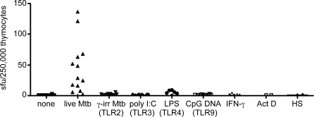
The control of Mycobacterium tuberculosis (Mtb) infection is heavily dependent on the adaptive Th1 cellular immune response. Paradoxically, optimal priming of the Th1 response requires activation of priming dendritic cells with Th1 cytokine IFN-gamma. At present, the innate cellular mechanisms required for the generation of an optimal Th1 T cell response remain poorly characterized. We hypothesized that innate Mtb-reactive T cells provide an early source of IFN-gamma to fully activate Mtb-exposed dendritic cells. Here, we report the identification of a novel population of Mtb-reactive CD4(-) alphabetaTCR(+) innate thymocytes. These cells are present at high frequencies, respond to Mtb-infected cells by producing IFN-gamma directly ex vivo, and display characteristics of effector memory T cells. This novel innate population of Mtb-reactive T cells will drive further investigation into the role of these cells in the containment of Mtb following infectious exposure. Furthermore, this is the first demonstration of a human innate pathogen-specific alphabetaTCR(+) T cell and is likely to inspire further investigation into innate T cells recognizing other important human pathogens.


Conflict of interest statement

Competing interests. The authors have declared that no competing interests exist.

Figures

Figure 1
Figure 1. Mtb-Reactive CD4 Thymocytes Are Present in the Human Thymus
(A) CD4-depleted thymocytes from thymocyte donor 11 (750,000/well) were incubated overnight with autologous DC (20,000/well) that were either infected with Mtb (moi of 50) or left uninfected. (B) Thymocytes (250,000 cells/well) from one thymocyte donor were incubated with MHC mismatched DC (50,000 cells/well) from 3 different donors. (C) Thymocytes from 4 individual donors were left unfractionated, or fractionated based on the positive or negative expression of CD4 using CD4 magnetic bead separation and incubated overnight with Mtb-infected DC (50,000/well). The dotted line represents the limit of detection of the assay. The lack of Mtb-reactivity from the CD4+ thymocytes fraction has been reproducible and repeated with over 10 donors. (D) CD4-depleted thymocytes (500,000/well) were titrated in a series of 2-fold dilutions and incubated with Mtb-infected DC (50,000/well). Frequencies were determined by linear regression analysis as described in Materials and Methods. In all experiments, IFN-γ production was assessed by ELISPOT. Mtb-reactive thymocytes (n = 18) ranged from 16.5 to 381 spot-forming units (sfu)/250,000 CD4 thymocytes with a mean of 115.9 ± SD 101.2.
Figure 2
Figure 2. The Majority of Mtb-Reactive Thymocytes Are αβTCR+ T Cells, While a Minority Express the γδTCR
(A) Thymocytes from 3 random donors were stained with the following fluorochrome-conjugated antibodies: CD4-PE, CD8-APC, γδTCR-FITC. CD4-negative subsets (γδTCR+; γδTCR CD8+; γδTCR CD8) were collected by FACS. Cell subsets were incubated with Mtb-infected DC or uninfected DC in an IFN-γ ELISPOT assay and the response to Mtb-infected DC is shown. No responses to uninfected DC were detected. (B) Unfractionated thymocytes (500,000 cells/well) were incubated with either uninfected or Mtb-infected DC (50,000 cells/well) and IFN-γ production was detected using the ICS assay. Cells were fixed and permeabilized and stained with fluorochrome-conjugated antibodies to label the following: γδTCR, αβTCR, IFN-γ, and CD3. All cells depicted in (B) are CD3+. Similar results were obtained from 3 separate donors.
Figure 3
Figure 3. Mtb-Reactive Thymocytes Display Molecules Associated with an Activated Effector Phenotype
(A) Unfractionated thymocytes (500,000 cells/well) and positive control PBMC (500,000 cells/well) were incubated with allogeneic DC (50,000 cells/well) infected with Mtb or left uninfected and IFN-γ assessed using the ICS assay. Non-specific binding by the mouse IgG1 PE isotype was not observed. The numbers in the graphs represent the percentage of IFN-γ-positive cells of unfractionated cells. Histograms represent expression of CD25, and CD8 on gated CD3+IFN-γ+ cells after stimulation with Mtb. (B) Histograms depicting granzyme and Bcl-2 expression from CD3+IFN-γ+-gated cells after stimulation with Mtb. Dotted line: isotype control antibody; Shaded histogram: granzyme or Bcl-2 expression on total CD3+ thymocytes; Bold line: granzyme or Bcl-2 expression on IFN-γ+ CD3+ cells (Mtb-reactive). Data are representative of a minimum of 4 thymocyte donors. (C) CD4-depleted thymocytes (500,000 cells/well) from 3 individuals were tested for their ability to secrete granzyme in an ELISPOT assay in response to DC (50,000 cells/well) that were uninfected or infected overnight with Mtb (moi 30).
Figure 4
Figure 4. Mtb-Reactive Thymocytes Are Stimulated by DCs Infected with Live Mtb but Not DCs Incubated with TLR Agonists
DC were incubated overnight with TLR agonists specific for TLR2 (γ-irradiated Mtb, moi equivalent of 500), TLR3 (poly I:C; 50 μg/ml), TLR4 (LPS; 100 ng/ml) TLR9 (CpG DNA; 6 μg/ml) or treated with IFN-γ (10 ng/ml), or Actinomycin D (10 μM) or heat shock-treated (42°C for 90 min) and were tested for their ability to stimulate DC to elicit IFN-γ production by thymocytes in comparison to live Mtb-infected DC (moi 30). Unfractionated thymocytes (250,000 cells/well) were incubated with the DC (50,000 cells/well) and tested for their ability to produce IFN-γ in an ELISPOT assay.
Figure 5
Figure 5. Mtb-Reactive Thymocytes Require Cell Contact with Mtb-Infected DC to Produce IFN-γ
CD4-depleted thymocytes (250,000 cells/well) were tested for their ability to produce IFN-γ in an ELISPOT assay in response to uninfected or Mtb-infected DC (50,000 cells/well) that were either placed directly in contact with the T cells (contact) on the ELISPOT membrane or placed in the upper wells of a 96-well Transwell plate (0.4-μm pore size) above the ELISPOT plate (transwell). Supernatants (100 μl) from uninfected or Mtb-infected DC were added directly to the T cells (supernatant). CD8+ T cell clones (15,000 cells/well) were used as positive controls for the response to Mtb-infected DC. Comparable results were obtained in 3 separate experiments.
Figure 6
Figure 6. Activation of Mtb-Reactive Thymocytes Is Proteasome Dependent but Is Not Blocked by the Pan–HLA-I Antibody W6/32
(A) DC were pretreated for 1 h with epoxomycin (1 μM). The DC were then infected with Mtb (moi 25). After 24 h, the drug was washed away and DC were fixed with cold paraformaldehyde (0.1%) for 5 min, washed twice in PBS, incubated for 2 h in media, and then washed three times. The DC (50,000 cells/well) were added to T cell clones (5000 cells/well) or CD4-depleted thymocytes (250,000 cell/well). Comparable results were obtained in 3 separate experiments. (B) Mtb-infected DC were incubated with the W6/32 antibody (2 μg/ml) or a mouse IgG2a isotype control (2 μg/ml) for 15 min before the addition of CD8+ T cell clones (10,000/well) or CD4-depleted thymocytes (250,000/well). IFN-γ production was evaluated by ELISPOT. The percent inhibition was calculated from the response to Mtb-infected DC in the presence of the W6/32 blocking antibody divided by the response to Mtb-infected DC in the presence of the isotype control.
Figure 7
Figure 7. Mtb-Reactive Cells Are Present in Cord Blood
Cord blood mononuclear cells (250,000 cells/well) were depleted of γδTCR+ cells using magnetic bead separation. γδTCR–depleted CBMC were incubated with autologous DC (50,000 cells/well) that were either Mtb-infected (moi = 30) of left uninfected. IFN-γ production was tested in an ELISPOT assay (n = 8; range, 0–80 sfu/250,000 γδTCR-depleted CBMC; mean = 26.75 ± SD 33.75).

References

    1. Maher D, Raviglione M. Global epidemiology of tuberculosis. Clin Chest Med. 2005;26:167–182. - PubMed
    1. Verver S, Warren RM, Munch Z, Richardson M, van der Spuy GD, et al. Proportion of tuberculosis transmission that takes place in households in a high-incidence area. Lancet. 2004;363:212–214. - PubMed
    1. Flynn JL. Immunology of tuberculosis and implications in vaccine development. Tuberculosis (Edinb) 2004;84:93–101. - PubMed
    1. Flynn JL, Chan J. Immunology of tuberculosis. Annu Rev Immunol. 2001;19:93–129. - PubMed
    1. Bhatt K, Hickman SP, Salgame P. Cutting edge: A new approach to modeling early lung immunity in murine tuberculosis. J Immunol. 2004;172:2748–2751. - PubMed

Publication types

MeSH terms

Substances