. 2008 Feb 8;4(2):e42.
doi: 10.1371/journal.ppat.0040042.

Structural insight into epitopes in the pregnancy-associated malaria protein VAR2CSA

Affiliations

Structural insight into epitopes in the pregnancy-associated malaria protein VAR2CSA

Pernille Andersen et al. PLoS Pathog. .

Abstract

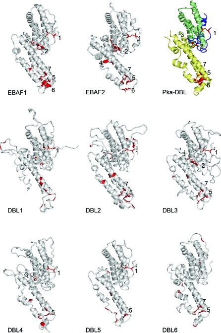
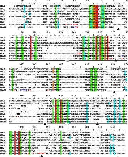
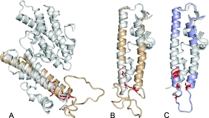
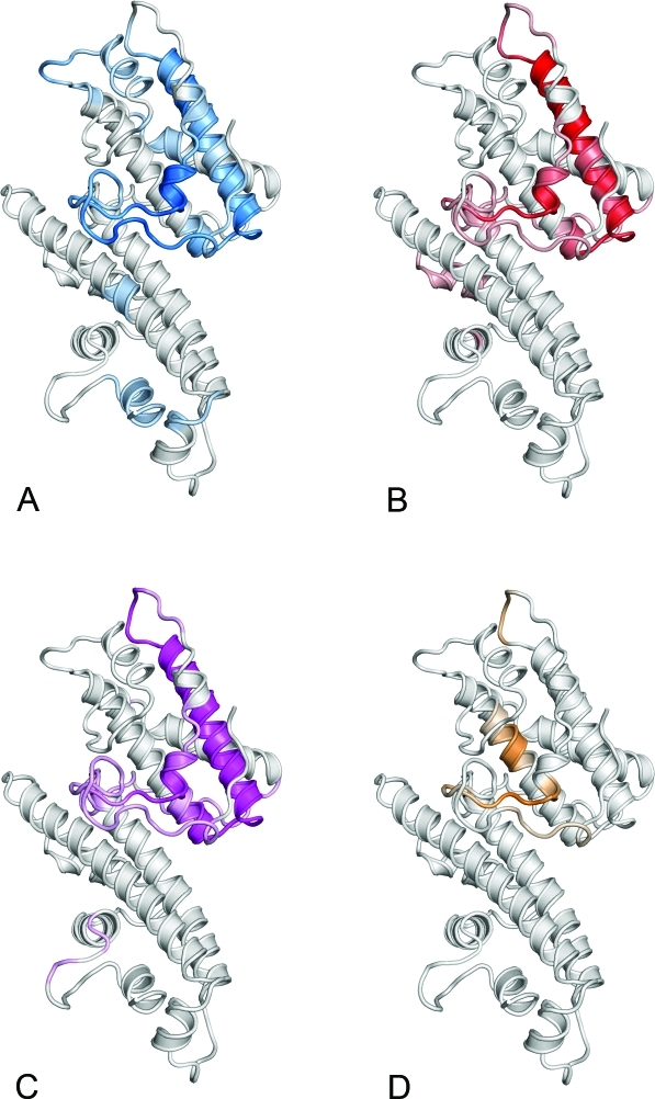
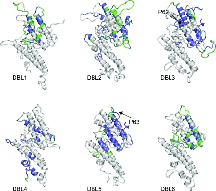
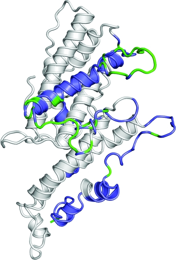
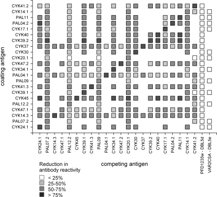
Pregnancy-associated malaria is caused by Plasmodium falciparum malaria parasites binding specifically to chondroitin sulfate A in the placenta. This sequestration of parasites is a major cause of low birth weight in infants and anemia in the mothers. VAR2CSA, a polymorphic multi-domain protein of the PfEMP1 family, is the main parasite ligand for CSA binding, and identification of protective antibody epitopes is essential for VAR2CSA vaccine development. Attempts to determine the crystallographic structures of VAR2CSA or its domains have not been successful yet. In this study, we propose 3D models for each of the VAR2CSA DBL domains and we show that regions in the fold of VAR2CSA inter-domain 2 and a PfEMP1 CIDR domain seem to be homologous to the EBA-175 and Pk alpha-DBL fold. This suggests that ID2 could be a functional domain. We also identify regions of VAR2CSA present on the surface of native VAR2CSA by comparing reactivity of plasma containing anti-VAR2CSA antibodies in peptide array experiments before and after incubation with native VAR2CSA. By this method we identify conserved VAR2CSA regions targeted by antibodies that react with the native molecule expressed on infected erythrocytes. By mapping the data onto the DBL models we present evidence suggesting that the S1+S2 DBL sub-domains are generally surface-exposed in most domains, whereas the S3 sub-domains are less exposed in native VAR2CSA. These results comprise an important step towards understanding the structure of VAR2CSA on the surface of CSA-binding infected erythrocytes.


Conflict of interest statement

Competing interests. The authors have declared that no competing interests exist.

Figures

Figure 1
Figure 1. Structure Models of the VAR2CSA DBL Domains
The experimental structures of the template EBA-175 and Pkα-DBL domains are shown in the first row, and DBL models are shown in the middle and last rows. Sub-domains 1–3 of Pkα-DBL (Pka-DBL) are colored in blue, green, and yellow, respectively. Cysteines in all domains are highlighted in red. Disulfide bonds in templates are numbered according to occurrence in the sequences and corresponding cysteine pairs in the models are numbered with respect to the template numbering.
Figure 2
Figure 2. A Structural Alignment of the Template Structures EBA-175 F1 and F2 and Pkα-DBL and the Six VAR2CSA DBL Models
Vertical color bars denote positions of stabilizing residues in template structures. Blue bars denote cysteines forming disulfide bonds, green bars denote buried hydrophobic residues, orange bars denote helix capping residues, and red bars denote buried polar/charged residues. The location of sub-domains S1–S3 are marked by horizontal black bars, and regions of missing structural information in the template structures are marked by black arrowheads. Residues involved in GAG binding of EBA-175 DBL domains or DARC receptor binding of Pkα-DBL are marked with red letters. EBA-175 residues involved in dimerization are marked in blue letters.
Figure 3
Figure 3. Modeled Regions of ID2 and CIDR Domains
The S3 domain of the template EBA-175 F2 is shown in white. Cysteines in the models are highlighted in red. (A) The model of the VAR2CSA ID2 domain superimposed in yellow on the EBA-175 F2. (B) Similar to (A) except showing only the S3 domain. (C) Model of a CIDR domain.
Figure 4
Figure 4. Mapping of Surface-Exposed Antibody-Reactive Regions onto the Model of the DBL6 Domain
The color intensity denotes the level of antibody depletion when VAR2CSA containing plasma is incubated with parasites expressing native VAR2CSA. The following plasma and parasite combinations were used for the depletion experiments. (A) Human plasma pool 1 depleted with 3D7 parasites. (B) Human plasma pool 2 depleted with 3D7 parasites. (C) Human plasma pool 2 depleted with FCR3 parasites. (D) Rabbit plasma pool depleted with 3D7 parasites.
Figure 5
Figure 5. Areas Predicted to be Targeted by Surface-Reactive Antibodies on the Six VAR2CSA DBL Domains
The highlighted regions were defined on the basis of the results of several experiments where anti-VAR2CSA antibody containing plasma was incubated with parasites expressing native VAR2CSA. Residues with consensus DVs > 1 which are conserved in sequences of VAR2CSA are highlighted in blue, and variable residues with consensus DVs>1 are highlighted in green. See text for details. The positions of DBL3 and DBL5 peptides (P62 and P63 respectively) used for affinity purifying IgG (see Figure 8) are marked with black arrows.
Figure 6
Figure 6. VAR2CSA DBL4 Regions Predicted to be Targeted by Surface-Reactive Antibodies
The plot is similar to Figure 4, except viewed from the reverse side.
Figure 7
Figure 7. A Matrix Showing the Level of Cross-Reactive Antibody Targets between 22 Placental VAR2CSA DBL3 Variants Produced in Baculovirus-Infected Insect Cells
Cross-reactivity was determined by competition ELISA using a Tanzanian female plasma pool with a high titer of VAR2CSA-specific antibodies. Responses with the same protein as the competing and coating antigen are shown in the diagonal. Blanks with no square are protein combinations not tested. The identity of the various DBL3 proteins corresponds to the nomenclature used in the published multiple sequence alignment [18]. Control proteins included a DBL5 VAR2CSA protein and a non-VAR2CSA DBL domain (PFD1235w).
Figure 8
Figure 8. Surface Reactivity (Fitch FL1) of Rabbit Antibodies Targeting Conserved VAR2CSA Regions
Rabbit antibodies were affinity purified on peptides (P62 and P63; see Figure 5 for details) and tested for reactivity with IE expressing VAR2CSA. The parasite lines FCR3CSA (A) and 3D7CSA (B) IE were used. The following antibodies were used: grey, IgG control; red, VAR2CSA-specific rabbit pool; blue, P63 (DBL5)-specific antibodies; green, P62 (DBL3)-specific antibodies. For FCR3, the number of cells staining positive with the VAR2CSA pool, P63 and P62 were 97%, 56%, and 48%, respectively. For 3D7, 85%, 7%, and 6% of the cells were positive.

Similar articles

Cited by

References

    1. Baruch DI, Pasloske BL, Singh HB, Bi X, Ma XC, et al. Cloning the P. falciparum gene encoding PfEMP1, a malarial variant antigen and adherence receptor on the surface of parasitized human erythrocytes. Cell. 1995;82:77–87. - PubMed
    1. Salanti A, Dahlback M, Turner L, Nielsen MA, Barfod L, et al. Evidence for the involvement of VAR2CSA in pregnancy-associated malaria. J Exp Med. 2004;200:1197–1203. - PMC - PubMed
    1. Weiss L. The spleen in malaria: The role of barrier cells. Immunol Lett. 1990;25:165–172. - PubMed
    1. Kraemer SM, Smith JD. A family affair: var genes, PfEMP1 binding, and malaria disease. Curr Opin Microbiol. 2006;9:374–380. - PubMed
    1. Fried M, Duffy PE. Adherence of Plasmodium falciparum to chondroitin sulfate A in the human placenta. Science. 1996;272:1502–1504. - PubMed

Publication types

MeSH terms

Substances