Review
. 2008:82:23-53.
doi: 10.1016/S0070-2153(07)00002-6.

The erythroblastic island

Affiliations
Review

The erythroblastic island

Deepa Manwani et al. Curr Top Dev Biol. 2008.

Abstract

Erythroblastic islands are specialized microenvironmental compartments within which definitive mammalian erythroblasts proliferate and differentiate. These islands consist of a central macrophage that extends cytoplasmic protrusions to a ring of surrounding erythroblasts. The interaction of cells within the erythroblastic island is essential for both early and late stages of erythroid maturation. It has been proposed that early in erythroid maturation the macrophages provide nutrients, proliferative and survival signals to the erythroblasts, and phagocytose extruded erythroblast nuclei at the conclusion of erythroid maturation. There is also accumulating evidence for the role of macrophages in promoting enucleation itself. The central macrophages are identified by their unique immunophenotypic signature. Their pronounced adhesive properties, ability for avid endocytosis, lack of respiratory bursts, and consequent release of toxic oxidative species, make them perfectly adapted to function as nurse cells. Both macrophages and erythroblasts display adhesive interactions that maintain island integrity, and elucidating these details is an area of intense interest and investigation. Such interactions enable regulatory feedback within islands via cross talk between cells and also trigger intracellular signaling pathways that regulate gene expression. An additional control mechanism for cellular growth within the erythroblastic islands is through the modulation of apoptosis via feedback loops between mature and immature erythroblasts and between macrophages and immature erythroblasts. The focus of this chapter is to outline the mechanisms by which erythroblastic islands aid erythropoiesis, review the historical data surrounding their discovery, and highlight important unanswered questions.


Figures

Figure 2.1
Figure 2.1
A three-dimensional scale model of a small volume of bone marrow from normal rat is shown. In this model, the large sphere is a megakaryocyte (m), and also shown are sinuses (S), macrophages (M), and the clusters of small spheres are the erythroblasts. This research was originally published in Blood (Mohandas and Prenant, 1978). © The American Society of Hematology.
Figure 2.2
Figure 2.2
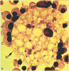
Characteristic appearance of human erythroblastic island. A Wright-Giemsa stained, cytocentrifuged preparation of cells obtained from erythroblasts generated by in vitro culture of peripheral blood-derived mononuclear cells using two-phase liquid culture system was analyzed. An erythroblastic island consisting of a central macrophage (M) surrounded by a ring of late erythroblasts (E) on day 12 of the second phase of a macrophage-containing culture is shown. This research was originally published in Blood (Hanspal et al., 1998). © The American Society of Hematology.
Figure 2.3
Figure 2.3
Reconstituted erythroblastic islands. Bright field (A), immunofluorescent standard (B), and confocal (C) micrographs of typical erythroblastic islands formed from single-cell suspensions of MacGreen mouse bone marrow. Macrophages from MacGreen mice express the macrophage colony-stimulating factor (M-CSF) receptor-green fluorescent protein transgene, thereby providing a useful macrophage identifier. Results from an assay for reforming islands from single-cell suspensions of freshly harvested mouse bone marrow are depicted. Adults 3–5 months of age were used. A single-cell suspension was prepared, then cells were incubated for carefully controlled times in media containing manganese. Islands and their cellular components were identified by three-color immunofluorescence microscopy. Immunofluorescent micrographs of islands show cells stained for erythroid-specific marker GPA (Ter119; red), macrophage marker M-CSF receptor GFP transgene expression (green), and DNA (Hoechst 33342; blue). Because surface expression of glycophorin A increases during terminal differentiation, the intensity of Ter119 staining served as an effective indicator of erythroblast stage. A faint blush of Ter119 fluorescence was present in early erythroblasts and increasing degrees of staining were observed in progressively more differentiated cells. The fluorescence intensity of Ter119 label varied among erythroblasts in an individual island, indicating that islands were composed of erythroblasts at various stages of differentiation. Young, multilobulated reticulocytes were present in many islands, again consistent with prior descriptions of erythroblastic islands formed in vivo. In the confocal image, some of the cells appear blurred because they are not in the plane of focus. However, macrophage staining is apparent in various regions of the island. Reticulocytes, arrowheads; macrophage, arrows; bars represent 10 μm. (D) Histogram shows number of erythroblastic islands formed from 1×10⁵ single cells; n=10. Results are shown as mean ± SD. This research was originally published in Blood (Lee et al., 2006). © The American Society of Hematology.
Figure 2.4
Figure 2.4
Scanning electron micrography of an erythroblastic island from rat bone marrow. Erythroblastic island after 2 hours in vitro culture examined by scanning electron microscopy (×5000). Top left insert: the same island seen with light microscopy (Giemsa ×1026). Note the two nuclear extrusions, one early (bottom) and the other almost complete (top left). This research was originally published in Blood Cells (Marcel Bessis et al., 1978). © Springer-Verlag.
Figure 2.5
Figure 2.5
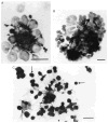
Analysis of erythroblastic islands after collagenase digestion and cluster purification. Direct cytocentrifuge preparations (A and B) or following adherence of cells to glass coverslip (C) are shown. (A) Small cluster stained with Ab F4/80, counterstained with hematoxylin, showing central macrophage surrounded by erythroblasts. Microscopic analysis at different depths of focus indicated that a single macrophage was present in such clusters. (B) Large cluster stained with Ab F4/80, counterstained with hematoxylin, showing four to five macrophages with processes ramifying among clustering cells. It has been proposed that this may represent several erythroblastic islands clumped together. (C) Central macrophage stained with Ab F4/80 after adhesion to glass coverslip. Note that delicate macrophage processes ramify extensively, establishing intimate contact with hematopoietic cells distal from the macrophage cell body (arrow) and appear to cradle hematopoietic cells. Counterstained with hematoxylin. Bar, 10 μm. This research was originally published in J. Exp Med. (Crocker and Gordon, 1985). © The Rockefeller University Press.

References

    1. Abboud S, Haile DJ. A novel mammalian iron-regulated protein involved in intracellular iron metabolism. J Biol Chem. 2000;275:19906–19912. - PubMed
    1. Allen TD, Dexter TM. Ultrastructural aspects of erythropoietic differentiation in long-term bone marrow culture. Differentiation. 1982;21:86–94. - PubMed
    1. Andrews NC, Schmidt PJ. Iron homeostasis. Annu Rev Physiol. 2006;69:69–85. - PubMed
    1. Arkin S, Naprstek B, Guarini L, Ferrone S, Lipton JM. Expression of intercellular adhesion molecule-1 (CD54) on hematopoietic progenitors. Blood. 1991;77:948–953. - PubMed
    1. Armeanu S, Buhring HJ, Reuss-Borst M, Muller CA, Klein G. E-cadherin is functionally involved in the maturation of the erythroid lineage. J Cell Biol. 1995;131:243–249. - PMC - PubMed

Publication types

LinkOut - more resources