. 2008 Feb 25;180(4):729-37.
doi: 10.1083/jcb.200707203. Epub 2008 Feb 18.

Dynamic behavior of GFP-CLIP-170 reveals fast protein turnover on microtubule plus ends

Affiliations

Dynamic behavior of GFP-CLIP-170 reveals fast protein turnover on microtubule plus ends

Katharina A Dragestein et al. J Cell Biol. .

Abstract

Microtubule (MT) plus end-tracking proteins (+TIPs) specifically recognize the ends of growing MTs. +TIPs are involved in diverse cellular processes such as cell division, cell migration, and cell polarity. Although +TIP tracking is important for these processes, the mechanisms underlying plus end specificity of mammalian +TIPs are not completely understood. Cytoplasmic linker protein 170 (CLIP-170), the prototype +TIP, was proposed to bind to MT ends with high affinity, possibly by copolymerization with tubulin, and to dissociate seconds later. However, using fluorescence-based approaches, we show that two +TIPs, CLIP-170 and end-binding protein 3 (EB3), turn over rapidly on MT ends. Diffusion of CLIP-170 and EB3 appears to be rate limiting for their binding to MT plus ends. We also report that the ends of growing MTs contain a surplus of sites to which CLIP-170 binds with relatively low affinity. We propose that the observed loss of fluorescent +TIPs at plus ends does not reflect the behavior of single molecules but is a result of overall structural changes of the MT end.


Figures
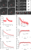
Figure 1.
Figure 1.
Fast FRAP analysis. (A–A′′′) Time-lapse imaging of COS-7 cells transiently expressing GFP–CLIP-170 (every 10th frame is shown). Two GFP–CLIP-170–labeled MT ends are indicated by arrows. In A, one of these is about to traverse a 210 × 210-nm ROI (red rectangles). After 2.25 s (A′′′), this MT end has traversed the ROI. (B) Fluorescence intensity in an ROI of 210 × 210 nm as a GFP–CLIP-170–labeled MT end traverses. (C and G) Mean fluorescence decay of nonbleached, GFP–CLIP-170-labeled (C) or EB3-GFP–labeled (G) MT ends (kdecay is indicated). (D–D′′′) COS-7 cells transiently expressing GFP–CLIP-170 were imaged as in A. An area of 256 × 3 pixels (indicated in D′) was bleached every 7.5 s, occasionally resulting in bleaching of MT end–bound GFP–CLIP-170. Rectangles 1–4 are shown enlarged underneath D–D′′′. Fluorescence intensity was measured in ROIs of 210 × 210 nm (red rectangles). (E) Fluorescence intensity of a bleached GFP–CLIP-170–labeled MT end (black arrow indicates bleach; black line indicates mean fluorescence decay). (F and H) Mean fluorescence recovery of GFP–CLIP-170 (F) and of EB3-GFP (H) on MT ends (krecovery is indicated).
Figure 2.
Figure 2.
Influence of temperature on GFP–CLIP-170 behavior. (A) GFP–CLIP-170 comets analyzed in stably expressing 3T3 cells (13 MT ends analyzed at 37°C, 19 at 32°C, and 14 at 27°C). (B) Mean fluorescence decay of GFP–CLIP-170–labeled MT ends in stably expressing 3T3 cells measured at 37°C (black; 13 ends analyzed) and at 27°C (red; 12 ends analyzed). A fit (see Materials and methods) yields a half-life of GFP–CLIP-170 fluorescence of 1.35 s at 37°C and 2.67 s at 27°C. (C) Mean fluorescence decay of GFP–CLIP-170–labeled MT ends in transiently transfected COS-7 cells at 37°C (black) and 27°C (red). See Table I for values. (D and E) Fast FRAP analysis at 27°C in MEFs transiently transfected with GFP–CLIP-170 (D) or GFP–CLIP-170XmnI (E). Fluorescent recovery was measured in the cytoplasm (blue) and on MT ends (red). For krecovery values, see Table I. Error bars indicate SEM.
Figure 3.
Figure 3.
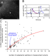
Analyzing GFP–CLIP-170 on MT ends with FCS. (A) Confocal image of a COS-7 cell transiently transfected with GFP–CLIP-170. The plus sign indicates the location of FCS measurement. Bar, 5 μm. (B) Intensity track of FCS measurement in a transfected COS-7 cell. Peaks of fluorescence are occasionally detected. The dotted red line indicates the exponential fluorescence decay; the bottom double-headed arrows indicate cytoplasmic fluorescence; the top double-headed arrows indicate peak fluorescence. (C) Comparison of the number of cytoplasmic and peak-bound GFP–CLIP-170 particles. Values as depicted in B were measured, and the number of particles was determined for 110 peaks. A scatter plot of these values is best approximated by the curve (indicated by the red line) Y = Ymax × (1 − e−kx).
Figure 4.
Figure 4.
Fast exchange model. (A) MT polymerization generates a large number of binding sites (orange ellipses), which disappear with single-order reaction kinetics. Thus, as time progresses, less binding sites are present within the depicted rectangle. (B) Dimeric CLIP-170 exchanges rapidly on binding sites irrespective of the position on the MT end. Several interactions with CLIP-170 molecules can occur during the lifetime of a binding site. The equilibrium between cytoplasmic and MT end–bound CLIP-170 (reaction a) might be determined by posttranslational modifications (reaction c), conformational changes, and/or protein–protein interactions. As we find CLIP-170 exchange on MT ends distal of sites of MT polymerization, copolymerization of CLIP-170 with tubulin (reaction b) does not explain the cometlike distribution of +TIPs. However, it is not excluded (hence the stippled arrow), and modified forms of CLIP-170 (indicated by the purple ellipses) might bind tubulin with higher affinity.

References

    1. Akhmanova, A., and C.C. Hoogenraad. 2005. Microtubule plus-end-tracking proteins: mechanisms and functions. Curr. Opin. Cell Biol. 17:47–54. - PubMed
    1. Akhmanova, A., A.-L. Mausset-Bonnefont, W. Van Cappellen, N. Keijzer, C.C. Hoogenraad, T. Stepanova, K. Drabek, J. van der Wees, M. Mommaas, J. Onderwater, et al. 2005. The microtubule plus end tracking protein CLIP-170 associates with the spermatid manchette and is essential for spermatogenesis. Genes Dev. 19:2501–2515. - PMC - PubMed
    1. Arnal, I., and R.H. Wade. 1995. How does taxol stabilize microtubules? Curr. Biol. 5:900–908. - PubMed
    1. Arnal, I., C. Heichette, G.S. Diamantopoulos, and D. Chretien. 2004. CLIP-170/tubulin-curved oligomers coassemble at microtubule ends and promote rescues. Curr. Biol. 14:2086–2095. - PubMed
    1. Charpilienne, A., M. Nejmeddine, M. Berois, N. Parez, E. Neumann, E. Hewat, G. Trugnan, and J. Cohen. 2001. Individual rotavirus-like particles containing 120 molecules of fluorescent protein are visible in living cells. J. Biol. Chem. 276:29361–29367. - PubMed

Publication types

MeSH terms

Substances