. 2008 Feb 25;180(4):739-46.
doi: 10.1083/jcb.200709161. Epub 2008 Feb 18.

Moesin and its activating kinase Slik are required for cortical stability and microtubule organization in mitotic cells

Affiliations

Moesin and its activating kinase Slik are required for cortical stability and microtubule organization in mitotic cells

Sébastien Carreno et al. J Cell Biol. .

Abstract

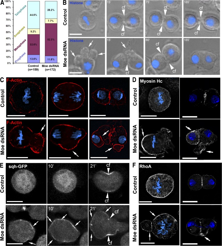
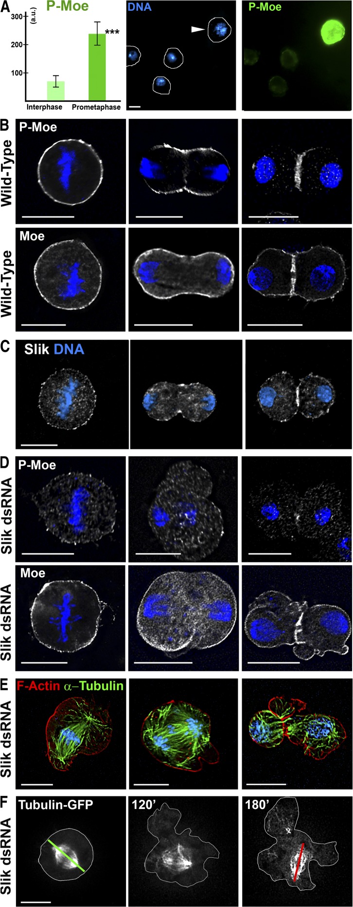
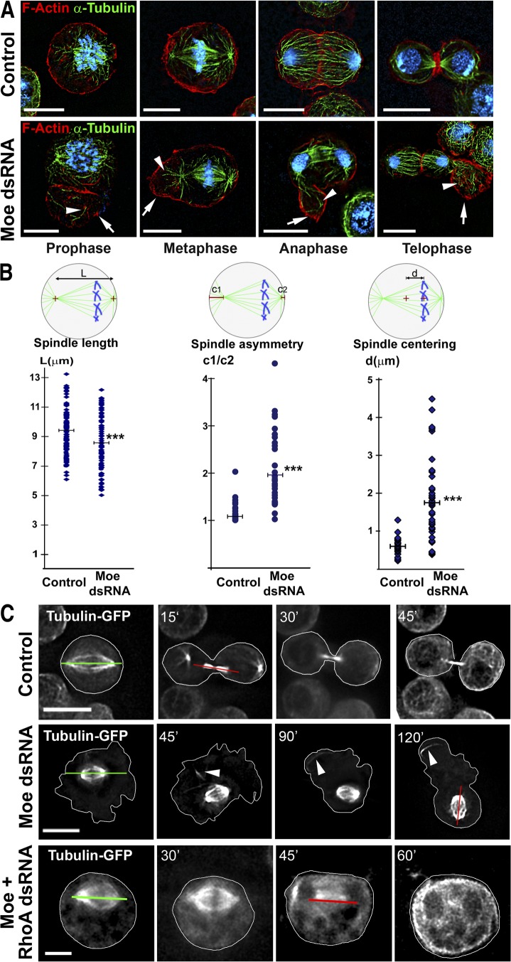
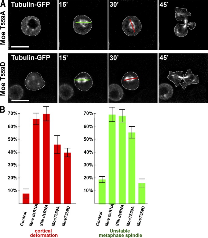
Cell division requires cell shape changes involving the localized reorganization of cortical actin, which must be tightly linked with chromosome segregation operated by the mitotic spindle. How this multistep process is coordinated remains poorly understood. In this study, we show that the actin/membrane linker moesin, the single ERM (ezrin, radixin, and moesin) protein in Drosophila melanogaster, is required to maintain cortical stability during mitosis. Mitosis onset is characterized by a burst of moesin activation mediated by a Slik kinase-dependent phosphorylation. Activated moesin homogenously localizes at the cortex in prometaphase and is progressively restricted at the equator in later stages. Lack of moesin or inhibition of its activation destabilized the cortex throughout mitosis, resulting in severe cortical deformations and abnormal distribution of actomyosin regulators. Inhibiting moesin activation also impaired microtubule organization and precluded stable positioning of the mitotic spindle. We propose that the spatiotemporal control of moesin activation at the mitotic cortex provides localized cues to coordinate cortical contractility and microtubule interactions during cell division.


Figures

Figure 1.
Figure 1.
Moesin controls cortex organization and contractility throughout mitosis. (A) Proportion of mitotic cells present in the respective steps of cell division, as deduced from fixed sample examination. Inactivation of moesin led to an increased proportion of cells in metaphase over the total number of mitotic cells. This increased proportion of metaphase indicates a delayed anaphase onset, which is also apparent in most time lapses. (B) Time-lapse frames of histone H2B–GFP (blue) S2 cells in control (top) or after moesin depletion (bottom) showing abnormal cortical protrusions (arrows) from pro/metaphase to ana/telophase. In this peculiar case, there was an eventual regression of the cytokinesis furrow even though the metaphase duration was not affected. cf, cleavage furrow. (C) F-actin (red) accumulated in ectopic cell bulges (arrows) in moesin-depleted cells throughout mitosis. DNA is in blue. (D) Consequences of moesin depletion on the distribution of myosin II heavy chain in metaphase and telophase. Myosin II heavy chain was irregularly localized (arrows) in the absence of moesin. (E) Actomyosin contractions were monitored in S2 cells expressing Sqh-GFP. Moesin depletion led to the chaotic distribution of contractile myosin (arrows) and contractions. (F) Consequences of moesin depletion on the distribution of active RhoA. RhoA was irregularly localized (arrows) in the absence of moesin. Bars, 10 μm.
Figure 2.
Figure 2.
The Slik kinase controls mitotic moesin function through regulation of its phosphorylation. (A) Relative levels of p-moesin (green) in prometaphase (arrowhead) and interphase cells. Error bars represent SD. ***, P < 0.005. (B and D) Distribution of total moesin or p-moesin during mitosis in control (B) or Slik-depleted cells (D). (C) Cells were immunolabeled to detect the Slik protein kinase, and DNA was counterstained with DAPI (blue). (E) Defects in cell shape and microtubule organization resulting from Slik depletion. α-Tubulin, green; F-actin, red; DNA, blue. Binucleated cells were observed in 5% of Slik-depleted cells (n = 500). (F) Time-lapse sequences of α-tubulin–GFP cells treated by Slik dsRNA. The red lines show maximal deviations of the spindle orientation from its initial position (green lines). Bars, 10 μm.
Figure 3.
Figure 3.
Moesin inactivation impairs mitotic spindle organization and positioning. (A) In the absence of moesin, abnormally long astral microtubules (arrowheads) contact the cortex at locations of actin-rich cell deformations (arrows). α-Tubulin, green; actin, red; DNA, blue. (B) Quantification of spindle defects in metaphase cells. L, mitotic spindle length (n = 100); c1/c2, ratio between distances of the centrosomes to their respective polar cortex (n = 25); d, distance between the geometrical center of the cell and the center of the spindle (n = 25). Each blue dot represents the calculated value for one cell. Horizontal bars represent the mean value of these data. ***, P < 0.005. (C) Time-lapse frames of S2 cells expressing α-tubulin–GFP treated with the indicated dsRNA. Arrowheads indicate astral microtubules associated with cortex bulges. The red lines show maximal deviations of the spindle orientation from its initial position (green lines). Bars, 10 μm.
Figure 4.
Figure 4.
Unregulated moesin phosphorylation affects organization of the cell cortex and microtubule spindle. (A) Selected frames of dividing α-tubulin–GFP cells expressing moesin-T559A or -T559D. The red lines show maximal deviations of the spindle orientation from its initial position (green lines). (B) Relative proportions of mitotic cells displaying cortical defects (left) and destabilization of the microtubule orientation (right) after depletion in moesin or Slik and after expression of the moesin-T559A or -T559D phosphomutant moesin forms. Error bars represent SD. Bars, 10 μm.

References

    1. Bement, W.M., H.A. Benink, and G. von Dassow. 2005. A microtubule-dependent zone of active RhoA during cleavage plane specification. J. Cell Biol. 170:91–101. - PMC - PubMed
    1. Bettencourt-Dias, M., R. Giet, R. Sinka, A. Mazumdar, W.G. Lock, F. Balloux, P.J. Zafiropoulos, S. Yamaguchi, S. Winter, R.W. Carthew, et al. 2004. Genome-wide survey of protein kinases required for cell cycle progression. Nature. 432:980–987. - PubMed
    1. Bjorklund, M., M. Taipale, M. Varjosalo, J. Saharinen, J. Lahdenpera, and J. Taipale. 2006. Identification of pathways regulating cell size and cell-cycle progression by RNAi. Nature. 439:1009–1013. - PubMed
    1. Bretscher, A., K. Edwards, and R.G. Fehon. 2002. ERM proteins and merlin: integrators at the cell cortex. Nat. Rev. Mol. Cell Biol. 3:586–599. - PubMed
    1. Canman, J.C., L.A. Cameron, P.S. Maddox, A. Straight, J.S. Tirnauer, T.J. Mitchison, G. Fang, T.M. Kapoor, and E.D. Salmon. 2003. Determining the position of the cell division plane. Nature. 424:1074–1078. - PubMed

Publication types

MeSH terms