

Morphogenesis of pancreatic cancer: role of pancreatic intraepithelial neoplasia (PanINs)
- PMID: 18283486
- PMCID: PMC2666329
- DOI: 10.1007/s00423-008-0282-x
Morphogenesis of pancreatic cancer: role of pancreatic intraepithelial neoplasia (PanINs)
Abstract
Introduction: Pancreatic ductal adenocarcinoma (i.e., pancreatic cancer) is an almost universally lethal disease. The identification of precursor lesions of pancreatic cancer provides an opportunity for early detection and potential therapeutic intervention before the development of invasive cancer.
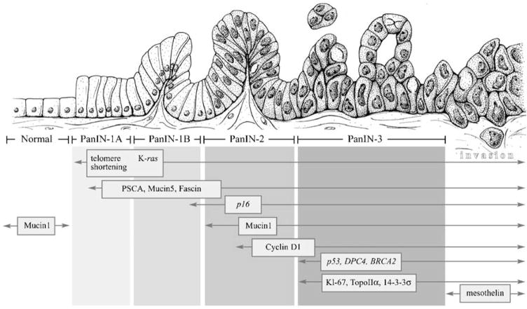
Discussion: It is now established that pancreatic cancers do not arise de novo but rather exhibit a sequential histological and genetic progression of precursor lesions culminating in frank, invasive neoplasia. Pancreatic intraepithelial neoplasia (PanIN) is the most common non-invasive precursor lesion of pancreatic cancer. The development of a consensus nomenclature scheme for PanINs has facilitated research into pancreatic cancer precursors and enabled standardization of results across institutions.
Conclusion: PanINs harbor many of the molecular alterations observed in invasive pancreatic cancer, confirming their status as true non-invasive precursor lesions. Recently developed genetically engineered mouse models of pancreatic cancer also demonstrate the stepwise PanIN progression model, underscoring the commonalities in pancreatic neoplasia between mouse and man.
Figures
References
-
- Parker JF, Florell SR, Alexander A, DiSario JA, Shami PJ, Leachman SA. Pancreatic carcinoma surveillance in patients with familial melanoma. Arch Dermatol. 2003;139:1019–1025. - PubMed
-
- Jemal A, Siegel R, Ward E, Murray T, Xu J, Smigal C, Thun MJ. Cancer statistics, 2006. CA Cancer J Clin. 2006;56:106–130. - PubMed
-
- American Cancer Society. Cancer facts & figures 2007. American Cancer Society; Atlanta: 2007. pp. 1–52.
-
- Sohn TA, Yeo CJ, Cameron JL, Koniaris L, Kaushal S, Abrams RA, Sauter PK, Coleman J, Hruban RH, Lillemoe KD. Resected adenocarcinoma of the pancreas-616 patients: results, outcomes, and prognostic indicators. J Gastrointest Surg. 2000;4:567–579. - PubMed
-
- Winter JM, Cameron JL, Campbell KA, Arnold MA, Chang DC, Coleman J, Hodgin MB, Sauter PK, Hruban RH, Riall TS, Schulick RD, Choti MA, Lillemoe KD, Yeo CJ. 1423 pancreaticoduodenectomies for pancreatic cancer: a single-institution experience. J Gastrointest Surg. 2006;10:1199–1210. - PubMed
Publication types
MeSH terms
Substances
Grants and funding
LinkOut - more resources
Full Text Sources
Other Literature Sources
Medical
Molecular Biology Databases
Miscellaneous
