. 2008 Dec;197(4):369-79.
doi: 10.1007/s00430-008-0081-6. Epub 2008 Feb 19.

Using distinct molecular signatures of human monocytes and dendritic cells to predict adjuvant activity and pyrogenicity of TLR agonists

Affiliations

Using distinct molecular signatures of human monocytes and dendritic cells to predict adjuvant activity and pyrogenicity of TLR agonists

Richard Kenmoe Kamgang et al. Med Microbiol Immunol. 2008 Dec.

Abstract

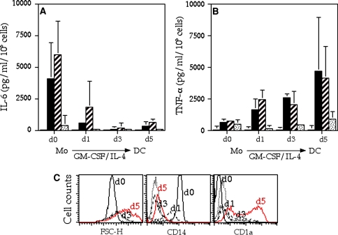
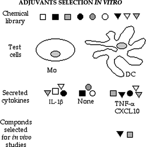
We present a systematic study that defines molecular profiles of adjuvanticity and pyrogenicity induced by agonists of human Toll-like receptor molecules in vitro. Using P(3)CSK(4), Lipid A and Poly I:C as model adjuvants we show that all three molecules enhance the expansion of IFNgamma(+)/CD4(+) T cells from their naïve precursors following priming with allogeneic DC in vitro. In contrast, co-culture of naive CD4(+) T cells with allogeneic monocytes and TLR2/TLR4 agonists only resulted in enhanced T cell proliferation. Distinct APC molecular signatures in response to each TLR agonist underline the dual effect observed on T cell responses. Using protein and gene expression assays, we show that TNF-alpha and CXCL10 represent DC-restricted molecular signatures of TLR2/TLR4 and TLR3 activation, respectively, in sharp contrast to IL-6 produced by monocytes upon stimulation with P(3)CSK(4) and Lipid A. Furthermore, although all TLR agonists are able to up-regulate proIL-1beta specific gene in both cell types, only monocyte activation with Lipid A results in detectable IL-1beta release. These molecular profiles, provide a simple screen to select new immune enhancers of human Th1 responses suitable for clinical application.


Figures

Fig. 1
Fig. 1
Effect of TLR2, TLR4 and TLR3 agonists on naive T cell activation. a CFSE-labeled CB-PBMC were cultured alone (white bars), or stimulated with allogeneic CD14+ Mo (hatched bars) or Mo-derived DC (black bars), in the presence of CSK4, P3CSK4, Lipid A, or Poly I:C, as indicated in the bottom line. T cell proliferation was assessed by measuring the frequency of CFSEdim/CD3+/CD4+ lymphocytes by FACS analysis. b Unlabeled CB-PBMC were stimulated as in a and assed for T cell differentiation by detection of intracellular IL-4 versus IFN-γ on gated CD3+/CD4+ lymphocytes; numbers indicate the percentage of cytokine-producing T cells. c Frequency of CD3+/CD4+/IFN-γ+ cells upon priming with allogeneic CD14+ Mo (white bars) or DC (black bars) in the presence of CSK4, P3CSK4, Lipid A or Poly I:C. Data represent mean results ± SD obtained from four independent experiments
Fig. 2
Fig. 2
Mo and DC respond distinctly to TLR2, TLR4 and TLR3 agonists in vitro. Freshly purified CD14+ cells (d0) and Mo undergoing DC differentiation (d1–d5) upon culture in GM-CSF/IL-4 conditioned medium, were stimulated with CSK4 (white bars), P3CSK4 (black bars), Lipid A (hatched bars) and Poly I:C (dotted bars). Secretion of IL-6 (a) and TNF-α (b) in culture supernatants was assessed by specific ELISA assays. Data are given as mean results ± SD obtained in four donors independently tested and are expressed in pg/ml/105 cells. c Time-dependent variation in cell size (FSC-H) and surface expression of CD14 and CD1a during Mo culture in GM-CSF/IL-4. Plots show profiles obtained with cells at days 0, 1, 3 and 5 of differentiation by FACS analysis. Results are representative of four experiments
Fig. 3
Fig. 3
Time-course analysis of cytokines release upon TLR2, TLR3 and TLR4 triggering on CD14+ Mo and DC in vitro. Freshly purified Mo (a, b) or Mo-derived DC (c, d) were stimulated, at 2 × 105 cells/ml, with CSK4 (open circle), P3CSK4 (filled circle), Lipid A (filled square) and Poly I:C (open square) and culture supernatants assessed for IL-6 (a, c) and TNF-α (b, d) secretion at different time points between 0.5 and 22 h. Representative results obtained in two out of three donors tested are shown
Fig. 4
Fig. 4
TLR3 is specifically expressed on DC and CXCL10 represents a cell-restricted signature of activation in response to Poly I:C. Freshly isolated CD14+ Mo were cultured in the presence of GM-CSF/IL-4 and analyzed by FACS staining or qRT-PCR at days (d) 0, 1, 3 and 5. a TLR2 surface expression in freshly isolated Mo (d0) and terminally differentiated DC (d5). In each plot dotted lines represent the background staining with an isotype-matched control antibody. b Quantitative gene expression analysis of TLR1, TLR2, TLR3 and TLR4 specific mRNA; data are expressed as Relative Quantity (R. Q.); cells at day 5 of culture were taken as calibrator (R. Q. = 1). Time-dependent induction of CCL2 and CXCL10 chemokines in response to CSK4 (white bars), P3CSK4 (black bars), Lipid A (hatched bars) and Poly I:C (dotted bars) in Mo undergoing DC differentiation in vitro is shown in c and d, respectively. Data are represented as described in Fig. 2
Fig. 5
Fig. 5
IL1β is a molecular marker of pyrogenicity. Freshly purified CD14+ cells (d0) and Mo undergoing DC differentiation (d1–d5) upon culture in GM-CSF/IL-4 conditioned medium, were stimulated with CSK4 (white bars), P3CSK4 (black bars), Lipid A (gray bars) and Poly I:C (dotted bars). Amounts of IL-1β a in culture supernatants, as assessed by specific ELISA assays are expressed in pg/ml/105 cells. Data are representative of three independent experiments. b Dose-dependent induction of IL-1β in Mo stimulated with CSK4 (open circle), P3CSK4 (filled circle) and Lipid A (filled triangle) at 104–1 ng/ml in threefolds serial dilutions. Experimental data obtained from one out of three donors independently tested are expressed in pg/ml and shown as mean results of triplicate wells
Fig. 6
Fig. 6
Prediction of adjuvant activity and safety of synthetic and natural compounds. a Correlation between amount of TNF-α released by DC and induction of Th1 responses in vitro upon stimulation with different natural and synthetic TLR-2 agonists. b Selection of putative safe adjuvants among a chemical library of 14 compounds including TLR-dependent and -independent stimuli, according to the capacity of each molecule to induce TNF-α secretion from activated DC (left panel), expansion of IFN-γ+/CD4+ T lymphocytes in MLR cultures (central panel) and IL-1β from CD14+ Mo in vitro (right panel). Cutoff for positive and negative selection are indicated by red and black arrows, respectively. Positively selected compounds are labeled in red
Fig. 7
Fig. 7
Schematic representation of an applicable in vitro system for adjuvants selection. In the proposed methods chemical compounds of unknown adjuvant activity are tested for their capacity to induce IL-1β release from human Mos (Mo) and TNF-α/CXCL10 secretion by Mo-derived dendritic cells (DC). Only chemicals positive for TNF-α/CXCL10 and negative for IL-1β are selected for in vivo studies

References

    1. {'text': '', 'ref_index': 1, 'ids': [{'type': 'DOI', 'value': '10.1038/ni1112', 'is_inner': False, 'url': 'https://doi.org/10.1038/ni1112'}, {'type': 'PubMed', 'value': '15454922', 'is_inner': True, 'url': 'https://pubmed.ncbi.nlm.nih.gov/15454922/'}]}
    2. Iwasaki A, Medzhitov R (2004) Toll-like receptor control of the adaptive immune responses. Nat Immunol 5:987–995 - PubMed
    1. {'text': '', 'ref_index': 1, 'ids': [{'type': 'DOI', 'value': '10.1146/annurev.immunol.21.120601.141126', 'is_inner': False, 'url': 'https://doi.org/10.1146/annurev.immunol.21.120601.141126'}, {'type': 'PubMed', 'value': '12524386', 'is_inner': True, 'url': 'https://pubmed.ncbi.nlm.nih.gov/12524386/'}]}
    2. Takeda K, Kaisho T, Akira S (2003) Toll-like receptors. Annu Rev Immunol 21:335–376 - PubMed
    1. {'text': '', 'ref_index': 1, 'ids': [{'type': 'PubMed', 'value': '12091878', 'is_inner': True, 'url': 'https://pubmed.ncbi.nlm.nih.gov/12091878/'}]}
    2. Alexopoulou L, Thomas V, Schnare M, Lobet Y, Anguita J, Schoen RT, Medzhitov R, Fikrig E, Flavell RA (2002) Hyporesponsiveness to vaccination with Borrelia burgdorferi OspA in humans and in TLR1- and TLR2-deficient mice. Nat Med 8:878–884 - PubMed
    1. {'text': '', 'ref_index': 1, 'ids': [{'type': 'DOI', 'value': '10.1038/35099560', 'is_inner': False, 'url': 'https://doi.org/10.1038/35099560'}, {'type': 'PubMed', 'value': '11607032', 'is_inner': True, 'url': 'https://pubmed.ncbi.nlm.nih.gov/11607032/'}]}
    2. Alexopoulou L, Holt AC, Medzhitov R, Flavell RA (2001) Recognition of double-stranded RNA and activation of NF-kappaB by Toll-like receptor 3. Nature 413:732–738 - PubMed
    1. {'text': '', 'ref_index': 1, 'ids': [{'type': 'DOI', 'value': '10.1073/pnas.040565397', 'is_inner': False, 'url': 'https://doi.org/10.1073/pnas.040565397'}, {'type': 'PMC', 'value': 'PMC15771', 'is_inner': False, 'url': 'https://pmc.ncbi.nlm.nih.gov/articles/PMC15771/'}, {'type': 'PubMed', 'value': '10681462', 'is_inner': True, 'url': 'https://pubmed.ncbi.nlm.nih.gov/10681462/'}]}
    2. Poltorak A, Ricciardi-Castagnoli P, Citterio S, Beutler B (2000) Physical contact between lipopolysaccharide and toll-like receptor 4 revealed by genetic complementation. Proc Natl Acad Sci USA 97:2163–2167 - PMC - PubMed

Publication types

MeSH terms