Review
. 2008 Apr;105(2):287-95.
doi: 10.1111/j.1471-4159.2008.05301.x. Epub 2008 Feb 14.

A role for glutamate in growth and invasion of primary brain tumors

Affiliations
Review

A role for glutamate in growth and invasion of primary brain tumors

Harald Sontheimer. J Neurochem. 2008 Apr.

Abstract

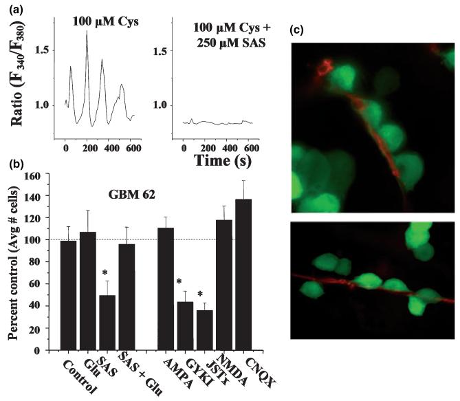
The vast majority of primary brain tumors derive from glial cells and are collectively called gliomas. While, they share some genetic mutations with other cancers, they do present with a unique biology and have developed adaptations to meet specific biological needs. Notably, glioma growth is physically restricted by the skull, and, unless normal brain cells are destroyed, tumors cannot expand. To overcome this challenge, glioma cells release glutamate which causes excitotoxic death to surrounding neurons, thereby vacating room for tumor expansion. The released glutamate also explains peritumoral seizures which are a common symptom early in the disease. Glutamate release occurs via system X(c), a cystine-glutamate exchanger that releases glutamate in exchange for cystine being imported for the synthesis of the cellular antioxidant GSH. It protects tumor cells from endogenously produced reactive oxygen and nitrogen species but also endows tumors with an enhanced resistance to radiation- and chemotherapy. Pre-clinical data demonstrates that pharmacological inhibition of system X(c) causes GSH depletion which slows tumor growth and curtails tumor invasion in vivo. An Food and Drug Administration approved drug candidate is currently being introduced into clinical trials for the treatment of malignant glioma.


Figures

Fig. 1
Fig. 1
Gliomas destroy normal brain as the tumor expands. Examples of patient images from four different patients that are presented with malignant gliomas, each illustrating the fact that normal brain tissue was displaced to accommodate the tumors expansion.
Fig. 2
Fig. 2
Gliomas release glutamate. (a) Gliomas in vivo do not express the GLT-1 glutamate transporter (green) while the surrounding astrocytes do. The GLT-1 staining demarcates the tumor boundaries. Red staining is propridium iodine to label cell nuclei. (b) Glioma cells release glutamate as determined by sampling medium in a 70-cm3 flask as a function of time. By contrast non-malignant astrocytes remove glutamate when challenged with 100 μM at the start. (c) Time-laps video microscopy shows pronounced neuronal cell death when hippocampal neurons are placed in the vicinity of glioma cells yet without touching them (sandwich culture). With permission from: (Ye and Sontheimer 1999).
Fig. 3
Fig. 3
Cystine-glutamate exchange in gliomas. (a) Glioma cell lines and acute biopsies highly express the catalytic (xCT) and regulatory (4F2hc) subunit of the system Xc cystine glutamate exchanger. (b) this transporter is responsible for the release of glutamate from the tumor into the surrounding brain. (c) In normal glia, glutamate released via system Xc is taken back up by the Na+-dependent GLT-1 transporter. (d) In gliomas, the loss of this reuptake via GLT-1 causes a buildup of glutamate and excitotoxic injury. (e) The main purpose of the system Xc transporter is to supply cysteine for the production of the cellular antioxidant GSH.
Fig. 4
Fig. 4
Inhibition of system Xc causes GSH depletion and reduces tumor size in vivo. (a) Sulfasalazine, which inhibits system Xc, causes a dose- and time-dependent reduction in GSH levels. (b) Intraperitoneal injection, twice daily, of 8 mg/kg sulfasalazine causes a 75% reduction in tumor volume in tumor bearing animals. (c) A representative comparison of control, saline injected to sulfasalazine treated animal at 30 days treatment. With permission from (Chung et al. 2005).
Fig. 5
Fig. 5
Glioma cells respond to glutamate as they invade. (a) Ca2+ imaging shows oscillatory changes in intracellular Ca2+ if cells are maintained in the presence of extracellular cystine (Cys), stimulating glutamate release via the cystine-glutamate exchanger. Ca2+ oscillations are abolished if cystine-glutamate exchange is inhibited with sulfasalazine (SAS). (b) Inhibition of glutamate release by SAS also inhibits Transwell migration, as does blockade of Ca2+ permeable AMPA receptors with GYKI or JSTx. Data represent normalized cell number as percentage of untreated control cells. Importantly SAS inhibition is overcome by exogenous addition of glutamate. (c) Glioma cells that express enhanced green fluorescent protein were imaged as they invaded cortical brain slices in situ. Cells typically migrate along blood vessel where they display ‘chain migration’ [a and c with permission from (Lyons et al. 2007)].

References

    1. Azizi SA, Miyamoto C. Principles of treatment of malignant gliomas in adults: an overview. J. Neurovirol. 1998;4:204–216. - PubMed
    1. Behrens PF, Langemann H, Strohschein R, Draeger J, Hennig J. Extracellular glutamate and other metabolites in and around RG2 rat glioma: an intracerebral microdialysis study. J. Neurooncol. 2000;47:11–22. - PubMed
    1. Biber K, Neumann H, Inoue K, Boddeke HW. Neuronal ‘On’ and ‘Off’ signals control microglia. Trends Neurosci. 2007;30:596–602. - PubMed
    1. Bordey A, Sontheimer H, Trouslard J. Muscarinic activation of BK channels induces membrane oscillations in glioma cells and leads to inhibition of cell migration. J. Membr. Biol. 2000;176:31–40. - PubMed
    1. Bryant JA, Finn RS, Slamon DJ, Cloughesy TF, Charles AC. EGF activates intracellular and intercellular calcium signaling by distinct pathways in tumor cells. Cancer Biol. Ther. 2004;3:1243–1249. - PubMed

Publication types

MeSH terms