. 2008 Feb 19:3:5.
doi: 10.1186/1749-8104-3-5.

Amplification of neural stem cell proliferation by intermediate progenitor cells in Drosophila brain development

Affiliations

Amplification of neural stem cell proliferation by intermediate progenitor cells in Drosophila brain development

Bruno C Bello et al. Neural Dev. .

Abstract

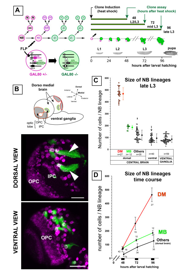
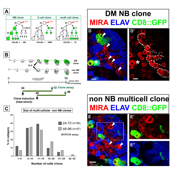
Background: In the mammalian brain, neural stem cells divide asymmetrically and often amplify the number of progeny they generate via symmetrically dividing intermediate progenitors. Here we investigate whether specific neural stem cell-like neuroblasts in the brain of Drosophila might also amplify neuronal proliferation by generating symmetrically dividing intermediate progenitors.

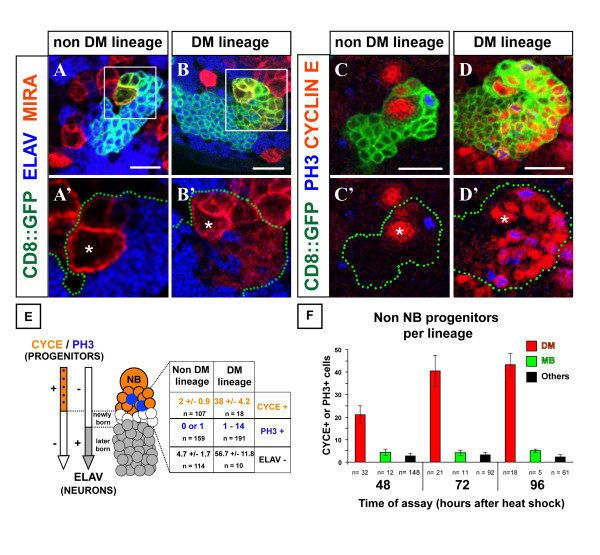
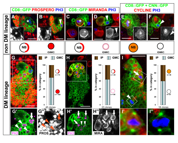
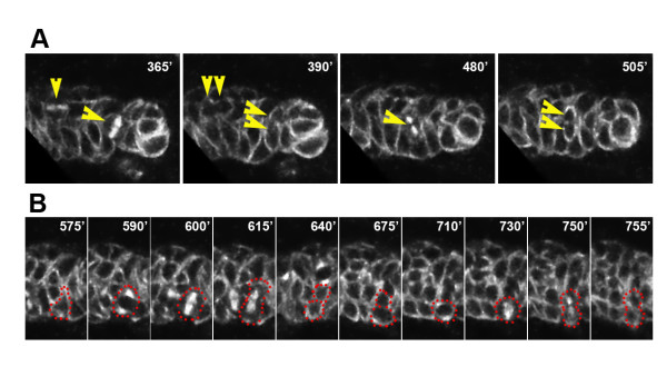
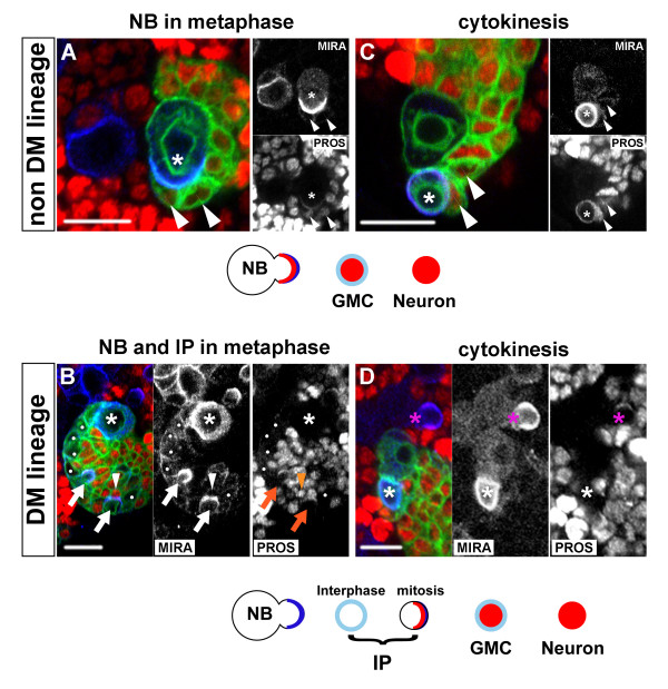
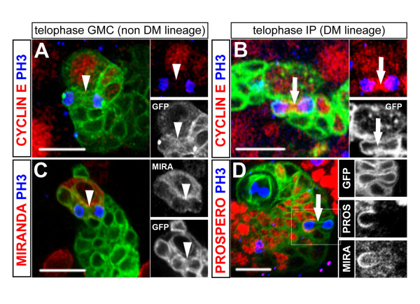
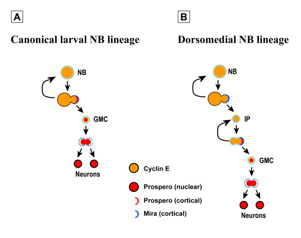
Results: Cell lineage-tracing and genetic marker analysis show that remarkably large neuroblast lineages exist in the dorsomedial larval brain of Drosophila. These lineages are generated by brain neuroblasts that divide asymmetrically to self renew but, unlike other brain neuroblasts, do not segregate the differentiating cell fate determinant Prospero to their smaller daughter cells. These daughter cells continue to express neuroblast-specific molecular markers and divide repeatedly to produce neural progeny, demonstrating that they are proliferating intermediate progenitors. The proliferative divisions of these intermediate progenitors have novel cellular and molecular features; they are morphologically symmetrical, but molecularly asymmetrical in that key differentiating cell fate determinants are segregated into only one of the two daughter cells.

Conclusion: Our findings provide cellular and molecular evidence for a new mode of neurogenesis in the larval brain of Drosophila that involves the amplification of neuroblast proliferation through intermediate progenitors. This type of neurogenesis bears remarkable similarities to neurogenesis in the mammalian brain, where neural stem cells as primary progenitors amplify the number of progeny they generate through generation of secondary progenitors. This suggests that key aspects of neural stem cell biology might be conserved in brain development of insects and mammals.


Figures

Figure 1
Figure 1
The DM brain NBs generate a large number of progeny during larval development.(a) Lineage labeling of a NB by MARCM. Left: schematic representation of a NB lineage in transgenic flies carrying a repressor transgene GAL80 distal to an FRT site in heterozygous (±) conditions. Ubiquitous expression of GAL80 under tubulin promoter control (pink) prevents GAL4-driven expression of the mCD8::GFP marker gene (green). Heat shock-induced FLP recombinase (FLP) at a given time point mediates the FRT site-specific mitotic recombination. Segregation of recombinant chromosomes at mitosis may result in the loss of the GAL80 repressor transgene in the NB daughter, which allows stable expression of the marker in this cell and its progeny. After several rounds of division such a positively labeled clone contains the NB, one or more GMCs and numerous post-mitotic neurons (N). Right: following random heat-shock induced NB recombination in newly hatched larvae, the size and composition of isolated NB lineages were examined at different time points during larval development. (b) NB clones were examined in all parts of the brain and ventral ganglia with the exception of optic lobes. The latter are easily recognizable in a single brain hemisphere by their lateral position and the high density of cells that express the progenitor marker Miranda (magenta, lower panels). On confocal images of brain hemispheres at low magnification (lower panels), GFP-labeled NB clones are easily identifiable by the presence of a large Miranda-positive NB and an associated cluster of clonal progeny. Unusually large clones could be identified in the dorsomedial part of the brain hemispheres (arrowheads). Anterior is to the top and lateral is to the left for each view. OPC and IPC, outer and inner proliferating centers, respectively. Scale bars: 50 μm. (c) The size of NB lineages was determined by counting cells in isolated clones plotted on the diagram according to their position in the nervous system (x axis). Each dot represents a clone with the mean ± standard deviation indicated by dots and error bars next to each group. DM, dorsomedial NB lineage; MB, mushroom body NB lineage; n, number of clones examined in each area. (d) Growth rate of different lineages examined at different time points after clone induction. Dots and bars represent the average size and standard deviation determined from the indicated number of clones.
Figure 2
Figure 2
The DM NBs generate an exceptional number of neuronal progenitors.(a-d') Confocal images of representative non-DM and DM lineages labeled with mCD8::GFP (membrane marker, green) in larval brains stained for the markers indicated. Each panel shows the most superficial area of a single NB clone viewed around the NB (asterisk) in the dorsal brain. The GFP channel is omitted for clarity in the lower panels and green dots outline the clones. Note that (a', b') show close up views of the areas boxed in (a, b). Progenitor cells in an NB lineage include the NB identifiable by its size (asterisk) and the most recently born cells in its associated progeny. These cells are found in close spatial proximity to the NB and are characterized by a weak level of cortical Miranda (red in a-b') and the absence of the neuronal marker ELAV (blue in a-b'). (c-d') NB-associated cells are unambiguously defined as progenitors by the expression of the cell cycle markers Cyclin E and/or PH3. (e) Quantification of various markers in NB clones at 96 h ALH underscores the high number of small progenitor cells among the progeny of the DM NBs. (f) DM NBs are always associated with the highest number of non-NB progenitors during larval development. Cell counts were performed on three types of clones recorded on the same samples for comparisons: DM, dorsomedial NB clones; MB, mushroom body NB clones; others, clones chosen at random in dorsal areas of the brain and not belonging by position and morphology to the other groups. In each case, the average number of progenitors is plotted with an error bar representing standard deviation. The number of clones examined is indicated bellow. Scale bars: 10 μm.
Figure 3
Figure 3
Molecular characterization of NB-like and GMC-like progenitors in the progeny of DM NBs. Confocal images of MARCM-labeled NB clones in the dorsal part of larval brains stained for the markers indicated on the top of the columns. Representative views of (a-f) non-DM lineages are used as a reference for (g-i") the DM lineages. Clones were labeled with CD8::GFP (membrane marker, green in all panels) and CNN::GFP (centrosomes visualized as bright green spots in e, f, i-i"). Proliferative cells are detected by anti-Cyclin (red in e, f, i-i') and anti-PH3 during mitosis (blue in all panels). In a non-DM NB clone, mitosis is restricted to two cell types: the NB and a single GMC in close proximity (a-f, asterisks and arrowheads, respectively). NBs show a unique pattern of polarized expression of Prospero and Miranda at the cell cortex during mitosis (a, c) and stable expression of Cyclin E throughout the cell cycle (e, mitosis; f, interphase). In contrast, the GMC is uniquely defined when engaged in mitosis (PH3 positive) by nuclear localization of Prospero (b, inset), weak uniform cortical localization of Miranda (d, inset) and lack of Cyclin E (f, inset). (g-i) In DM clones many progenitors other than the NB are identified as PH3-positive nuclei. These cells show patterns of marker expression usually found in mitotic NBs (IP; arrows) or mitotic GMCs (arrowheads). Lower panels show close up views of the areas boxed in (g-i). The two types of mitotic progenitors can be detected simultaneously in a single DM lineage (images) and are found at a comparable ratio when quantified in multiple clones using the three independent markers (histograms). IP, small NB-associated intermediate progenitor with NB-like marker expression. Scale bars: 10 μm (a-f) or 15 μm (g-i).
Figure 4
Figure 4
Live imaging of multiple and repeated division of DM NB daughter cells in MARCM-labeled clones. Frames from time-lapse recordings of a DM clone labeled with CD8::GFP and tau::GFP in larval brain cultured over 13 hours. The large NB, not visible in these frames, divided twice during this time period (Additional data file 1). The time is indicated in minutes relative to the start of the recording. (a) Multiple divisions of small NB-associated cells may be ongoing simultaneously in the clone and each gives rise to two daughter cells of equal size (single and double arrowheads at following intervals). (b) A single NB daughter cell may undergo several rounds of division. Shown are two consecutive divisions of a cell outlined with dots. Following a first symmetric division (575'–675'), the lower daughter cell underwent a second division (710'–755') while its sibling did not divide further during the recording.
Figure 5
Figure 5
Clonal expansion of IPs analyzed by MARCM.(a) Schematic representation of the different types of MARCM clones that can be recovered following FLP-mediated recombination in a NB (red arrow) and segregation of homozygous GAL80 chromosomes into one of its two daughter cells (green). A multicellular clone lacking the NB (right panel) reveals the ability of the IP daughter cell to undergo several rounds of division. Not shown are FLP-mediated recombination events in the GMC or in the IP that give rise to multicellular clones only in the latter case. Recombination in the GMC gives a single labeled cell. (b) Top: schematic organization of multicellular GFP-labeled clones (green) after time-controlled recombination (heat-shocked FLP, black arrows) in two developing NB lineages. Bottom: unlike NB clones (upper lineage), IP clones were identified as GFP-labeled cell clusters lacking the large Miranda-positive NB and pushed away from this founder cell by proliferation (non-NB clone, lower lineage). The size and composition of clonal progenies were examined 48 hours after two independent heat-induced recombination events. (c) Size distribution of multicellular non-NB clones generated by recombination at 24 h (light grey bars) or 48 h (dark grey bars) ALH and assayed 48 hours later. The similar histogram profile reveals the comparable mitotic potential of progenitors present in the DM lineage at 24 or 48 h ALH. (d, d') Representative confocal image of NB clones induced at 48 h ALH and examined at 96 h in a dissected brain stained for the markers indicated (dorsal view, lateral to the left, anterior to the top). DM NBs are identifiable in the most medial row of large cells (arrowheads) by their association to a large cluster of Miranda-positive progenitors (various DM lineages are outlined by dots in (d); the GFP channel was omitted for clarity). The GFP-labeled progeny of a single DM NB follows the orientation of the Miranda-positive cell cluster. A typical non-DM NB clone is found on the lateral site of the brain (asterisk). This single large NB is associated with a few Miranda-positive GMCs. (e) Representative IP clone of four cells among the presumptive progeny of the nearest DM NB (arrowheads); same scale and conditions as in (d). A magnification of the area boxed in (e) is shown in (e', e"), with one channel omitted for clarity. The cells in the clone have undetectable level of Miranda (red) and all express the neuronal marker ELAV (blue). Scale bars: 15 μm.
Figure 6
Figure 6
Asymmetrically dividing DM NBs do not express Prospero. Confocal images of NB divisions in a canonical NB lineage (top panels) compared to a DM lineage (bottom panels). Shown are representative CD8::GFP-labeled clones (green), seen around the NB in late larval brains stained for Miranda (MIRA, blue) and Prospero (PROS, red). Single channels are also shown in gray scale for better contrast. (a, b) Miranda forms cortical crescents at metaphase in both non-DM and DM NBs (asterisk). (c, d) Following asymmetric division, Miranda segregates into the small daughter cell and remains associated at high levels at the cortex soon after cytokinesis (the small newborn daughter cell is marked by an asterisk). Prospero co-localizes with Miranda in the dividing non-DM NBs (a, c, asterisks) and is nuclear in the oldest GMCs, which retain a low level of Miranda at the cortex (a, c, arrowheads), and in all other post-mitotic cells in the clone. In the DM NBs, Prospero is undetectable during mitosis (b, d, asterisks). (Note in (d) a canonical NB outside the clone (magenta asterisk) that shows co-localization of Miranda and Prospero and serves as internal control.) Recently born NB daughter cells show weak uniform cortical Miranda and lack Prospero (white dots in b). Polarized cortical Miranda during mitosis identifies these cells as IPs (b, arrows) and co-localization with Prospero is once again observed in these cells (b, insets). Cells with GMC-like (arrowheads) or neuronal expression of the markers are also observed as in canonical non-NB lineages. Scale bars: 10 μm.
Figure 7
Figure 7
Unequal segregation of Prospero/Miranda during symmetric division of IPs. Confocal images of representative CD8::GFP labeled clones (green) in (a, c) canonical non-DM or (b, d) DM lineages. Shown are mitotic figures of small NB-associated cells at anaphase/telophase, visualized by anti- PH3 staining of DNA (blue). Separate channels are also shown in insets for better contrast. The outline of the plasma membrane stained by CD8::GFP shows that both the GMC (a, c, arrowheads) and the IP (b, d, arrows) divide symmetrically and give rise to daughter cells of similar sizes. The dividing IP is identified by NB-like expression of Cyclin E during mitosis (b) while GMC division lacks Cyclin E expression at this phase of the cell cycle (a). In the mitotic GMC, Miranda distributes equally to both daughter cells (c, inset) while Prospero is nuclear (see Figure 3b). In IP division, Prospero and Miranda co-segregate to only one of the two daughter cells (d, insets). Scale bars: 10 μm.
Figure 8
Figure 8
Model for a transient amplifying progenitor cells in DM NB lineages.(a) In the canonical model of asymmetric NB division, a single neurogenic division of the small GMC progenitor cell produces two neurons (N) at each round of NB division. Unequal partitioning of Prospero promotes neurogenic division by inhibiting self-renewing factors in the GMC. (b) The DM NB divides asymmetrically without Prospero, which enables the small daughter cell to retain self-renewing potential and to behave as an IP. In this cell, expression of Prospero and unknown polarization cues re-established the asymmetric segregation of fate determinants and the generation of the neurogenic progenitor GMC. This novel mode of neurogenesis increases the number of post-mitotic neurons that individual NBs in the dorsomedial brain can generate at each round of divisions.

References

    1. Gotz M, Huttner WB. The cell biology of neurogenesis. Nat Rev Mol Cell Biol. 2005;6:777–788. doi: 10.1038/nrm1739. - DOI - PubMed
    1. Merkle FT, Alvarez-Buylla A. Neural stem cells in mammalian development. Curr Opin Cell Biol. 2006;18:704–709. doi: 10.1016/j.ceb.2006.09.008. - DOI - PubMed
    1. Kriegstein A, Noctor S, Martinez-Cerdeno V. Patterns of neural stem and progenitor cell division may underlie evolutionary cortical expansion. Nat Rev Neurosci. 2006;7:883–890. doi: 10.1038/nrn2008. - DOI - PubMed
    1. Morrison SJ, Kimble J. Asymmetric and symmetric stem-cell divisions in development and cancer. Nature. 2006;441:1068–1074. doi: 10.1038/nature04956. - DOI - PubMed
    1. Al-Hajj M, Clarke MF. Self-renewal and solid tumor stem cells. Oncogene. 2004;23:7274–7282. doi: 10.1038/sj.onc.1207947. - DOI - PubMed

Publication types

MeSH terms

LinkOut - more resources