Expression and protein localisation of IGF2 in the marsupial placenta
- PMID: 18284703
- PMCID: PMC2276195
- DOI: 10.1186/1471-213X-8-17
Expression and protein localisation of IGF2 in the marsupial placenta
Abstract
Background: In eutherian mammals, genomic imprinting is critical for normal placentation and embryo survival. Insulin-like growth factor 2 (IGF2) is imprinted in the placenta of both eutherians and marsupials, but its function, or that of any imprinted gene, has not been investigated in any marsupial. This study examines the role of IGF2 in the yolk sac placenta of the tammar wallaby, Macropus eugenii.
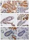
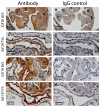
Results: IGF2 mRNA and protein were produced in the marsupial placenta. Both IGF2 receptors were present in the placenta, and presumably mediate IGF2 mitogenic actions. IGF2 mRNA levels were highest in the vascular region of the yolk sac placenta. IGF2 increased vascular endothelial growth factor expression in placental explant cultures, suggesting that IGF2 promotes vascularisation of the yolk sac.
Conclusion: This is the first demonstration of a physiological role for any imprinted gene in marsupial placentation. The conserved imprinting of IGF2 in this marsupial and in all eutherian species so far investigated, but not in monotremes, suggests that imprinting of this gene may have originated in the placenta of the therian ancestor.
Figures


Similar articles
-
Insulin is imprinted in the placenta of the marsupial, Macropus eugenii.Dev Biol. 2007 Sep 15;309(2):317-28. doi: 10.1016/j.ydbio.2007.07.025. Epub 2007 Jul 27. Dev Biol. 2007. PMID: 17706631
-
Characterisation of marsupial PHLDA2 reveals eutherian specific acquisition of imprinting.BMC Evol Biol. 2011 Aug 19;11:244. doi: 10.1186/1471-2148-11-244. BMC Evol Biol. 2011. PMID: 21854573 Free PMC article.
-
Genomic imprinting of IGF2, p57(KIP2) and PEG1/MEST in a marsupial, the tammar wallaby.Mech Dev. 2005 Feb;122(2):213-22. doi: 10.1016/j.mod.2004.10.003. Mech Dev. 2005. PMID: 15652708
-
The origin and evolution of genomic imprinting and viviparity in mammals.Philos Trans R Soc Lond B Biol Sci. 2013 Jan 5;368(1609):20120151. doi: 10.1098/rstb.2012.0151. Philos Trans R Soc Lond B Biol Sci. 2013. PMID: 23166401 Free PMC article. Review.
-
Genomic imprinting in marsupial placentation.Reproduction. 2008 Nov;136(5):523-31. doi: 10.1530/REP-08-0264. Epub 2008 Sep 19. Reproduction. 2008. PMID: 18805821 Review.
Cited by
-
Marsupial genome sequences: providing insight into evolution and disease.Scientifica (Cairo). 2012;2012:543176. doi: 10.6064/2012/543176. Epub 2012 Nov 25. Scientifica (Cairo). 2012. PMID: 24278712 Free PMC article. Review.
-
Placental expression of pituitary hormones is an ancestral feature of therian mammals.Evodevo. 2011 Aug 19;2:16. doi: 10.1186/2041-9139-2-16. Evodevo. 2011. PMID: 21854600 Free PMC article.
-
Selected imprinting of INS in the marsupial.Epigenetics Chromatin. 2012 Aug 28;5(1):14. doi: 10.1186/1756-8935-5-14. Epigenetics Chromatin. 2012. PMID: 22929229 Free PMC article.
-
Placentation in Marsupials.Adv Anat Embryol Cell Biol. 2021;234:41-60. doi: 10.1007/978-3-030-77360-1_4. Adv Anat Embryol Cell Biol. 2021. PMID: 34694477
-
Promoter-specific expression and imprint status of marsupial IGF2.PLoS One. 2012;7(7):e41690. doi: 10.1371/journal.pone.0041690. Epub 2012 Jul 25. PLoS One. 2012. PMID: 22848567 Free PMC article.
References
-
- Mossman H. Comparative morphogenesis of the fetal membranes and accessory uterine structures. Contrib Embryol. 1937;26:129–246. - PubMed
-
- Amoroso EC. Placentation. In: Parkes AS, editor. Marshall's Physiology of Reproduction. Vol. 2. London: Longmans Green; 1952. pp. 127–311.
-
- Luckett WP. Ontogeny of amniote fetal membranes and their application to phylogeny. In: Hecht M, Goody P, Hecht B, editor. Major Patterns in Vertebrate Evolution. New York: Plenum Press; 1977. pp. 439–516.
Publication types
MeSH terms
Substances
LinkOut - more resources
Full Text Sources
Miscellaneous

