. 2008 Apr 18;283(16):10408-14.
doi: 10.1074/jbc.M710474200. Epub 2008 Feb 19.

Specific oxidized phospholipids inhibit scavenger receptor bi-mediated selective uptake of cholesteryl esters

Affiliations

Specific oxidized phospholipids inhibit scavenger receptor bi-mediated selective uptake of cholesteryl esters

Mohammad Z Ashraf et al. J Biol Chem. .

Abstract

We have recently demonstrated that specific oxidized phospholipids (oxPC(CD36)) accumulate at sites of oxidative stress in vivo such as within atherosclerotic lesions, hyperlipidemic plasma, and plasma with low high-density lipoprotein levels. oxPC(CD36) serve as high affinity ligands for the scavenger receptor CD36, mediate uptake of oxidized low density lipoprotein by macrophages, and promote a pro-thrombotic state via platelet scavenger receptor CD36. We now report that oxPC(CD36) represent ligands for another member of the scavenger receptor class B, type I (SR-BI). oxPC(CD36) prevent binding to SR-BI of its physiological ligand, high density lipoprotein, because of the close proximity of the binding sites for these two ligands on SR-BI. Furthermore, oxPC(CD36) interfere with SR-BI-mediated selective uptake of cholesteryl esters in hepatocytes. Thus, oxidative stress and accumulation of specific oxidized phospholipids in plasma may have an inhibitory effect on reverse cholesterol transport.


Figures

FIGURE 1.
FIGURE 1.
Binding of NO2-LDL and oxPCCD36 to SR-BI-transfected cells. a, 125I-LDL or various modified 125I-labeled lipoproteins (5 μg/ml) were incubated with SR-BI-transfected CHO cells (ldlA7-SR-BI) or control vector-transfected CHO cells (ldlA7). Cellular binding of lipoproteins was subsequently determined as described under “Experimental Procedures.” (Cu2+oxLDL, copper oxidized LDL). *, p < 0.001 for comparison versus LDL. b, 125I-labeled NO2-LDL was incubated with SR-BI-expressing cells either in the absence (N.A., no addition) or presence of the indicated competitors. Anti-SR-BI, anti-SR-BI antibody; N.I. IgG, nonimmune IgG. *, p < 0.001 for comparison versus control (no addition). c, small unilamellar vesicles were generated containing tracer levels of [3H]DPPC (25 μCi/mg vesicles) and consisting either of the unoxidized parent phospholipid alone (PAPC), phospholipid oxidized by formula image system (NO2-PAPC), or an equimolar mixture of parent phospholipid and the indicated synthetic oxidized phospholipids (oxPCCD36). Cells were incubated with phospholipid vesicles, and cell-associated radioactivity was quantified. *, p < 0.001 for comparison versus PAPC; #, p < 0.001 for comparison versus KDdiA-PC and versus KDdiA-PC + nonimmune (N.I.) IgG.
FIGURE 2.
FIGURE 2.
NO2-LDL and oxPCCD36 compete for the binding of HDL to SR-BI. a, 125I-HDL (5 μg/ml) was incubated with ldlA7-SR-BI cells or vector control cells, and bound 125I-HDL was quantified. The concentrations of competitors used were 200 μg of protein/ml for lipoproteins, 40 μg of lipid/ml for vesicles, and 20 μg/ml for antibody. Anti-SR-BI, anti-SR-BI antibody; N.I. IgG, nonimmune IgG.*, p < 0.001 for comparison versus control (N.A., no addition). b, 125I-HDL (5 μg/ml) was incubated with GST-SR-BI-(144–205) protein beads, either in the absence (no addition) or presence of 20-fold excess unlabeled indicated competitors in PBS/BSA. Beads were repeatedly washed with PBS to remove unbound lipoproteins, and bound radioactivity was then quantified. *, p < 0.001 for comparison versus control (no addition). c, GST-SR-BI-(144–205) protein beads were incubated with small unilamellar vesicles consisting of PAPC, NO2-PAPC, or the indicated oxPCCD36 with tracer levels of [3H]DPPC (25 μCi/mg vesicles) (10 μg/ml), washed repeatedly with PBS, and assayed for bead-associated radioactivity. *, p < 0.001 for comparison versus PAPC. #, p < 0.001 for comparison versus KDdiA-PC. d, concentration dependence of 125I-HDL binding to SR-BI-overexpressing HEK-293T cell and GST-SR-BI-(144–205) protein. Indicated concentrations of 125I-HDL were incubated with either cells or GST-SR-BI-(144–205) beads at 25 °C for 3 h and then washed, and bound radioactivity was quantified. Specific 125I-HDL binding was determined by subtracting background binding to control vector-transfected cells or GST beads alone, respectively.
FIGURE 3.
FIGURE 3.
Binding of NO2-LDL and oxPCCD36 to Hep G2 cells. a, 125I-NO2-LDL (5 μg/ml) was incubated with Hep G2 cells either in the absence (N.A., no addition) or presence of the indicated competitors at 4 °C for 3 h in the appropriate media. Cellular binding of lipoproteins was subsequently determined as described under “Experimental Procedures.” Anti-SR-BI, anti-SR-BI antibody; N.I. IgG, nonimmune IgG. *, p < 0.001 for comparison versus control (no addition). b, 125I-HDL (5 μg/ml) was incubated with Hep G2 cells either in the absence (no addition) or presence of the indicated competitors. *, p < 0.001 for comparison versus control (no addition). c, small unilamellar vesicles were generated as described in Fig. 1, and Hep G2 cells were incubated with phospholipid vesicles (10 μg/mg of cell protein), following 3 h of incubation at 4 °C and washed, and cell-associated radioactivity was quantified. *, p < 0.001 for comparison versus PAPC. #, p < 0.001 for comparison versus KDdiA-PC and versus KDdiA-PC + nonimmune IgG.
FIGURE 4.
FIGURE 4.
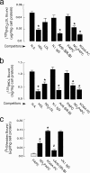
oxPCCD36 interfere with SR-BI-mediated selective cholesteryl ester uptake. Hep G2 cells were incubated with either [3H]COE HDL (a) or 125I-HDL (b) at a concentration 10 μg/ml for 5 h 37 °C, with indicated additions, and then cells were washed with warm PBS and chased for another 30 min in the presence of 100 μg/ml unlabeled HDL. a, cells were lysed; cell-associated radioactivity was quantified and [3H]COE HDL association determined. b, 125I-HDL uptake was determined as described under “Experimental Procedures.” c, selective uptake was calculated from data in a and b as described under “Experimental Procedures.” *, p < 0.001 for comparison versus control.

Similar articles

Cited by

References

    1. Arai, T., Wang, N., Bezouevski, M., Welch, C., and Tall, A. R. (1999) J. Biol. Chem. 274 2366–2371 - PubMed
    1. Kozarsky, K. F., Donahee, M. H., Glick, J. M., Krieger, M., and Rader, D. J. (2000) Arterioscler. Thromb. Vasc. Biol. 20 721–727 - PubMed
    1. Ueda, Y., Gong, E., Royer, L., Cooper, P. N., Francone, O. L., and Rubin, E. M. (2000) J. Biol. Chem. 275 20368–20373 - PubMed
    1. Huszar, D., Varban, M. L., Rinninger, F., Feeley, R., Arai, T., Fairchild-Huntress, V., Donovan, M. J., and Tall, A. R. (2000) Arterioscler. Thromb. Vasc. Biol. 20 1068–1073 - PubMed
    1. Acton, S. L., Scherer, P. E., Lodish, H. F., and Krieger, M. (1994) J. Biol. Chem. 269 21003–21009 - PubMed

Publication types