. 2008 May;56(5):495-509.
doi: 10.1369/jhc.2008.950592. Epub 2008 Feb 18.

Murine mCLCA6 is an integral apical membrane protein of non-goblet cell enterocytes and co-localizes with the cystic fibrosis transmembrane conductance regulator

Affiliations

Murine mCLCA6 is an integral apical membrane protein of non-goblet cell enterocytes and co-localizes with the cystic fibrosis transmembrane conductance regulator

Melanie K Bothe et al. J Histochem Cytochem. 2008 May.

Abstract

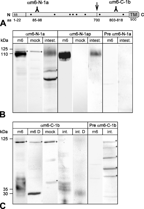
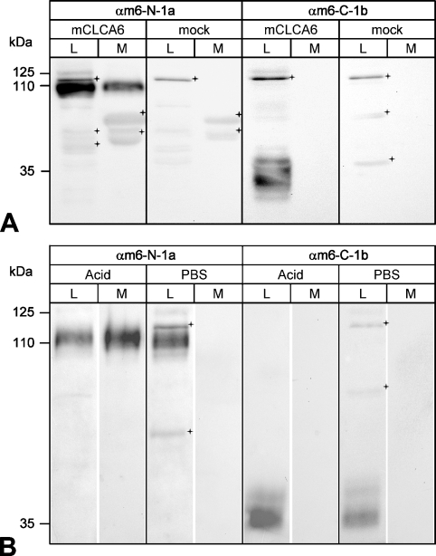
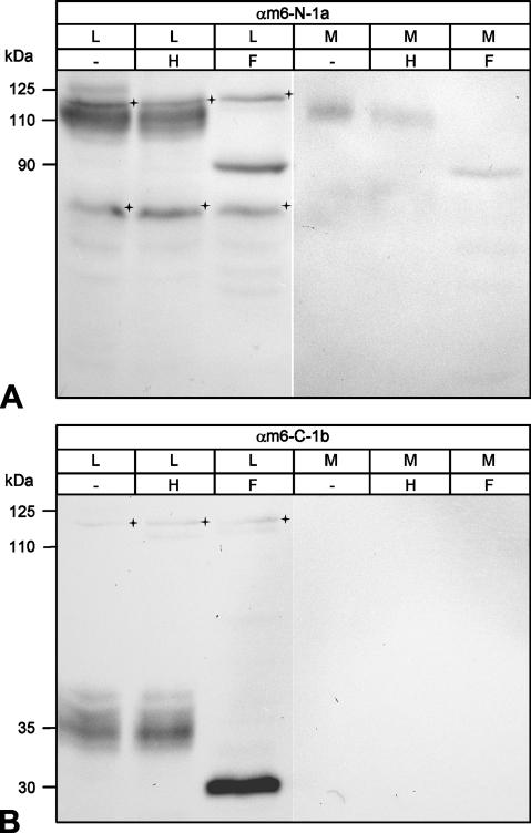
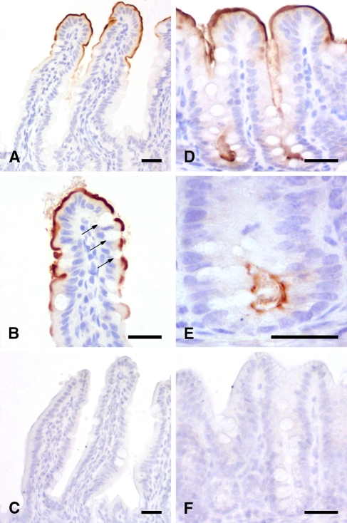
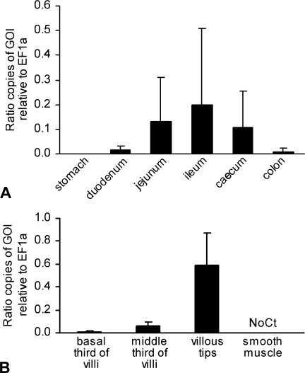
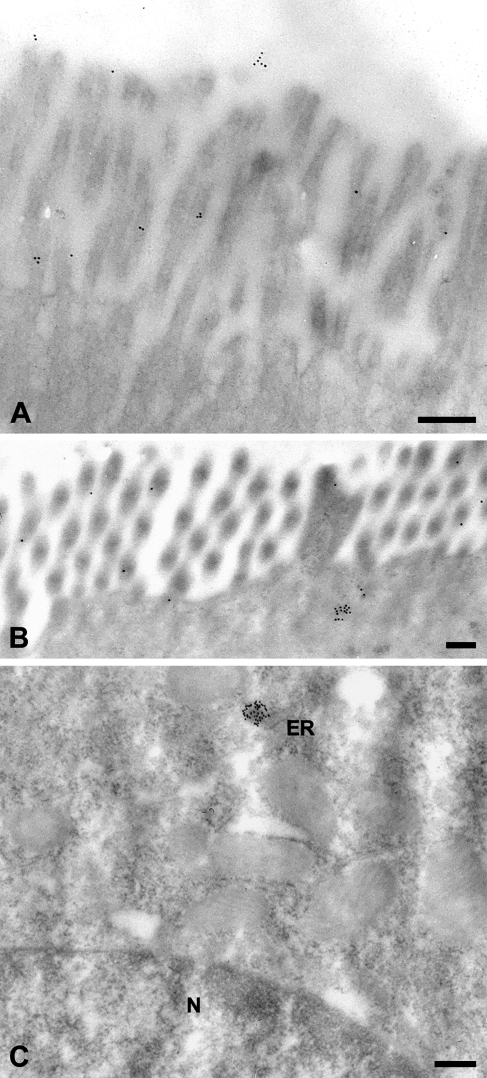
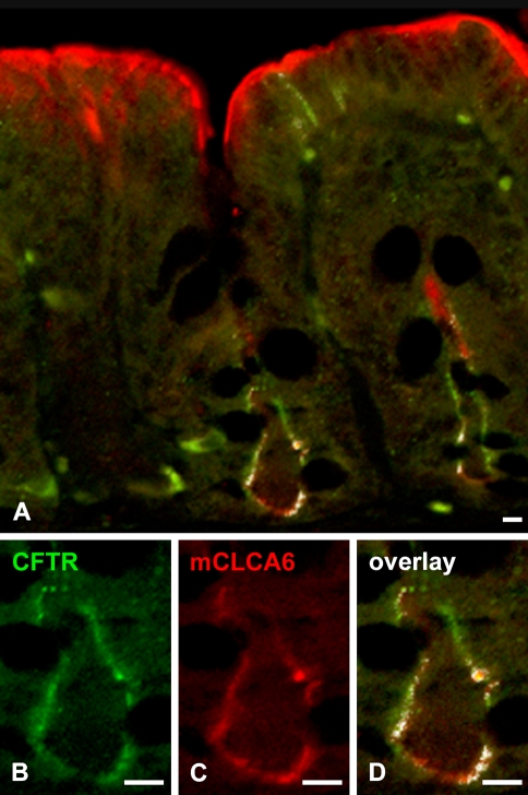
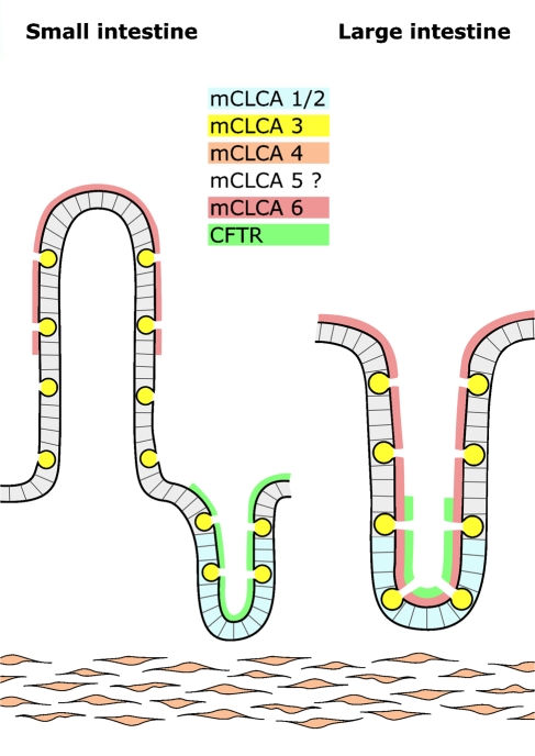
The CLCA family of proteins consists of a growing number of structurally and functionally diverse members with distinct expression patterns in different tissues. Several CLCA homologs have been implicated in diseases with secretory dysfunctions in the respiratory and intestinal tracts. Here we present biochemical protein characterization and details on the cellular and subcellular expression pattern of the murine mCLCA6 using specific antibodies directed against the amino- and carboxy-terminal cleavage products of mCLCA6. Computational and biochemical characterizations revealed protein processing and structural elements shared with hCLCA2 including anchorage in the apical cell membrane by a transmembrane domain in the carboxy-terminal subunit. A systematic light- and electron-microscopic immunolocalization found mCLCA6 to be associated with the microvilli of non-goblet cell enterocytes in the murine small and large intestine but in no other tissues. The expression pattern was confirmed by quantitative RT-PCR following laser-capture microdissection of relevant tissues. Confocal laser scanning microscopy colocalized the mCLCA6 protein with the cystic fibrosis transmembrane conductance regulator CFTR at the apical surface of colonic crypt cells. Together with previously published functional data, the results support a direct or indirect role of mCLCA6 in transepithelial anion conductance in the mouse intestine.


Figures

Figure 1
Figure 1
Generation of antibodies against mCLCA6. (A) Antisera were raised against synthetic peptides corresponding to amino acid (aa) sequences within the amino-(N)-terminal cleavage product (αm6-N-1a) and the carboxy-(C)-terminal cleavage product (αm6-C-1b). Hydrophobicity analyses predicted a signal sequence (ss) at the amino-terminus and a transmembrane domain (TM) at the end of the carboxy-terminal cleavage product. Dots indicate consensus sites for N-linked glycosylation. Arrow shows putative cleavage site. (B) When lysates of heterologously mCLCA6-transfected HEK293 cells were immunoblotted with αm6-N-1a, two specific proteins were detected of 125 and 110 kDa in size, respectively (Lane 1). Lysates of intestinal tissues contained only the 110-kDa protein (Lane 3). Preimmune serum from the same rabbit failed to detect any protein in the same lysates (Lanes 7 and 8). In addition to specific detection of mCLCA6, the unpurified immune serum αm6-N-1a reacted unspecifically with an additional protein of ∼115 kDa in transfected HEK293 cells (Lane 2) and two proteins of ∼70 and 60 kDa (crosses) in the intestinal lysate (Lane 4). However, these proteins were not detected by the immunopurified serum αm6-N-1ap. (C) Anti-carboxy-terminal antibody αm6-C-1b also detected the 125-kDa primary translation product only in overexpressing HEK293 cells (Lane 1), not in intestinal tissue lysates (Lanes 4 and 5). The various protein species of ∼35 kDa were reduced by deglycosylation with PNGase F to a single protein of ∼30 kDa in size (Lane 2, m6 D = cell lysate, deglycosylated; Lane 5, int. D = intestinal lysate, deglycosylated). Vector alone transfected cells served as controls (Lane 3, mock). All antibodies were diluted at 1:500. Crosses = unspecific background staining.
Figure 2
Figure 2
Cellular processing of mCLCA6. (A) Immunoblotting analyses identified both the amino- and the carboxy-terminal cleavage products of mCLCA6 in the cell lysate (L, Lanes 1 and 5) but only the N-terminal subunit in the conditioned medium (M, Lane 2). HEK293 cells transiently transfected with mCLCA6 were analyzed by immunoblotting using antibodies αm6-N-1a (1:500) or αm6-C-1b (1:500), respectively. For analysis of the conditioned medium, a medium change was performed 42 hr after transfection followed by harvest and analysis of the medium 6 hr later. Vector alone transfected cells served as controls (mock, Lanes 7 and 8). Crosses = unspecific background staining. (B) When cells were incubated at pH 2.5, only the N-terminal cleavage product of mCLCA6 was partially released into the supernatant. Forty eight hr after transfection, HEK293 cells transiently transfected with mCLCA6 were incubated with NaCl, pH 2.5 (pH adjusted with acetic acid) to release non-covalently associated proteins from the plasma membrane. Treatment with PBS served as control. Antibodies αm6-N-1a and αm6-C-1b were diluted at 1:500. Crosses = unspecific background staining.
Figure 3
Figure 3
Glycosylation patterns of the amino- and carboxy-terminal cleavage products. (A) Deglycosylation experiments identified the N-terminal cleavage product of mCLCA6 as a complex glycosylated mature protein form that has passed the Golgi apparatus. Aliquots of cell lysate (L) and supernatant (M) of HEK293 cells transiently transfected with mCLCA6 were treated with endo H (H), PNGase F (F), or not treated (−). The N-terminal cleavage product of mCLCA6 was resistant to endo H (Lane 2) but was reduced in size to ∼90 kDa when treated with PNGase F (Lane 3). Crosses = unspecific background staining. (B) The carboxy-terminal cleavage product in the cell lysate (L) was also resistant to endo H (Lane 2) and was reduced in size to appear 30 kDa when treated with PNGase F (Lane 3). Crosses = unspecific background staining.
Figure 4
Figure 4
mCLCA6 is located at the apical surface of non-goblet cell enterocytes. Immunohistochemical analyses localized mCLCA6 at the apical surface of non-goblet cell enterocytes at the villous tips of the jejunum (A,B) and in the crypts of the colon (D,E). Goblet cells stained negative (arrows in B). Tissue sections were incubated with αm6-N-1ap (diluted 1:20,000 in A,B,D,E) or the respective preimmune serum (diluted 1:20,000 in C,F). Color was developed using DAB as substrate (brown) with hematoxylin as counterstain (blue). Bar = 20 μm.
Figure 5
Figure 5
Quantification of mCLCA6 mRNA expression in the murine intestine using quantitative RT-PCR. (A) Relative copy numbers of mCLCA6 mRNA increased from the duodenum to the ileum and thereafter decreased to the colon, confirming the staining intensities observed using immunohistochemical protein detection. Bars = standard deviations. (B) Laser capture microdissection was used to isolate different segments along the crypt–villus axis of the jejunal villi prior to quantitative RT-PCR. Results confirmed increasing expression levels in the villous tips (compare with Figure 4A). Expression levels are given in ratios of gene of interest (GOI) relative to the copy numbers of the housekeeping gene EF1α (n=5 C57BL/6 mice). No expression was detected in jejunal smooth muscle (No Ct).
Figure 6
Figure 6
Immune electron microscopic localization of the mCLCA6 protein. On the ultrastructural level, the mCLCA6 protein was localized to the brush border of enterocytes (A,B) in small clusters underneath the terminal actin web, in small clusters associated with the endoplasmic reticulum near the nucleus (C), and in small numbers in the lumen of the intestine (A). Tissue sections were incubated with αm6-N-1ap (diluted 1:12,000). Incubation with αm6-C-1bp (1:50) resulted in the same staining pattern except for the lumen, which was negative for the carboxy-terminal cleavage product. Incubation without primary antibody served as negative control. ER, endoplasmic reticulum; N, nucleus. Bar = 200 nm.
Figure 7
Figure 7
Colocalization of mCLCA6 and CFTR in apical plasma membranes of non-goblet cell enterocytes in crypts of the large intestine. The mCLCA6 protein (red) was visualized along the apical plasma membrane of non-goblet cell enterocytes both at the intestinal lumen and in the crypts (A,C). In contrast, the CFTR protein was only detected at the apical plasma membrane of enterocytes in the deep crypt areas (green in A,B) where it was clearly colocalized with the mCLCA6 protein (A,C). (A,D) Overlay of both signals. (A,B) CFTR staining using antibody R3195 diluted 1:25 with FITC-labeled secondary antibody. (A,C) mCLCA6 staining using antibody αm6-N-1ap diluted 1:100 with LRSC-labeled secondary antibody. Bar = 10 μm.
Figure 8
Figure 8
Different structural and functional niches are occupied by distinct CLCA homologs in the murine intestine. The mCLCA1 and mCLCA2 protein (blue) are expressed in basal crypt epithelia of the small and large intestine (Gruber et al. 1998b; Leverkoehne et al. 2006), mCLCA3 (yellow) in secretory vesicles of goblet cells (Romio et al. 1999; Leverkoehne and Gruber 2002), mCLCA4 (orange) in smooth muscle cells (Elble et al. 2002), and mCLCA6 (red) on the apical surface of non-goblet cell enterocytes in the small intestinal villi (left panel) and throughout the large intestinal crypts (right panel). The cell type expressing mCLCA5 in the mouse intestine is so far unknown (Evans et al. 2004). CFTR chloride channel (green) has previously been localized to the apical surface of crypt enterocytes (Ameen et al. 2000a) where it is coexpressed with mCLCA6 in the large intestine.

References

    1. Abdel-Ghany M, Cheng HC, Elble RC, Lin H, DiBiasio J, Pauli BU (2003) The interacting binding domains of the β4 integrin and calcium-activated chloride channels (CLCAs) in metastasis. J Biol Chem 278:49406–49416 - PubMed
    1. Abdel-Ghany M, Cheng HC, Elble RC, Pauli BU (2001) The breast cancer β4 integrin and endothelial human CLCA2 mediate lung metastasis. J Biol Chem 276:25438–25446 - PubMed
    1. Agnel M, Vermat T, Culouscou JM (1999) Identification of three novel members of the calcium-dependent chloride channel (CaCC) family predominantly expressed in the digestive tract and trachea. FEBS Lett 455:295–301 - PubMed
    1. Al-Jumaily M, Kozlenkov A, Mechaly I, Fichard A, Matha V, Scamps F, Valmier J, et al. (2007) Expression of three distinct families of calcium-activated chloride channel genes in the mouse dorsal root ganglion. Neurosci Bull 23:293–299 - PMC - PubMed
    1. Ameen N, Alexis J, Salas P (2000a) Cellular localization of the cystic fibrosis transmembrane conductance regulator in mouse intestinal tract. Histochem Cell Biol 114:69–75 - PubMed

Publication types

MeSH terms