. 2008 May;28(9):3058-69.
doi: 10.1128/MCB.02025-07. Epub 2008 Feb 19.

A glycine-arginine domain in control of the human MRE11 DNA repair protein

Affiliations

A glycine-arginine domain in control of the human MRE11 DNA repair protein

Ugo Déry et al. Mol Cell Biol. 2008 May.

Abstract

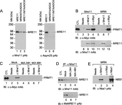
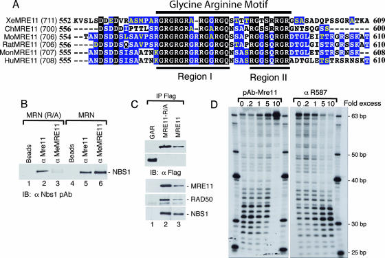
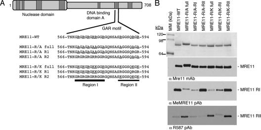
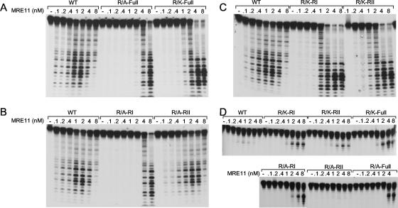
Human MRE11 is a key enzyme in DNA double-strand break repair and genome stability. Human MRE11 bears a glycine-arginine-rich (GAR) motif that is conserved among multicellular eukaryotic species. We investigated how this motif influences MRE11 function. Human MRE11 alone or a complex of MRE11, RAD50, and NBS1 (MRN) was methylated in insect cells, suggesting that this modification is conserved during evolution. We demonstrate that PRMT1 interacts with MRE11 but not with the MRN complex, suggesting that MRE11 arginine methylation occurs prior to the binding of NBS1 and RAD50. Moreover, the first six methylated arginines are essential for the regulation of MRE11 DNA binding and nuclease activity. The inhibition of arginine methylation leads to a reduction in MRE11 and RAD51 focus formation on a unique double-strand break in vivo. Furthermore, the MRE11-methylated GAR domain is sufficient for its targeting to DNA damage foci and colocalization with gamma-H2AX. These studies highlight an important role for the GAR domain in regulating MRE11 function at the biochemical and cellular levels during DNA double-strand break repair.


Figures

FIG. 1.
FIG. 1.
MRE11 is methylated in insect cells and interacts with PRMT1. (A) SF9 cells were infected with MRE11 (lanes 3 and 6) or MRE11, RAD50, and NBS1 baculoviruses (lanes 2 and 5) and treated with methyltransferase inhibitors MTA and ADOX for 24 h (lanes 2 to 3 and 5 to 6). Lanes 1 and 4, purified MRE11-His6. MM, molecular mass; PAb, polyclonal antibody. (B) Interactions between MRE11 and PRMT1. SF9 cells were coinfected with MRE11 and c-Myc-PRMT1 baculoviruses. Immunoprecipitations were conducted using beads alone (lanes 2, 5, 8, and 11), anti-MRE11 (lanes 3, 6, 9, and 12), or anti-c-Myc (lanes 4, 7, 10, and 13) and visualized by Western blotting as indicated. Lane 1, whole-cell extract from Sf9 cells infected with c-Myc PRMT1 baculovirus. CTL, control; IB, immunoblot; mAb, monoclonal antibody. (C) Treatment of Sf9 cells coinfected with MRE11 and PRMT1 viruses with methyltransferase inhibitors MTA and ADOX disrupt MRE11-PRMT1 interaction. SF9 cells were coinfected with MRE11 and c-Myc-PRMT1 baculoviruses, and immunoprecipitations were conducted using beads alone (lanes 2, 4, and 6) or anti-MRE11 (lanes 3, 5, and 7) and visualized by Western blotting, as indicated. Lane 1, whole-cell extract from Sf9 cells infected with c-Myc PRMT1 baculovirus. (D) Control figure showing that MTA and ADOX treatment inhibit MRE11 methylation in the presence of PRMT1. SF9 cells coinfected with MRE11 and PRMT1 baculoviruses were mock treated (lanes 2 and 5) or treated with MTA and ADOX (M/A) for 24 h or 48 h (lanes 3 and 6 and 4 and 7, respectively). Lane 1, purified MRE11-His6. MRE11 was immunoprecipitated with anti-MRE11 polyclonal antibody and revealed with anti-MRE11 and anti-MeMRE11 polyclonal antibody, which detects methylated arginines in MRE11. IP, immunoprecipitate. (E) PRMT1 does not interact with the MRN complex. SF9 cells were coinfected with MRE11, RAD50, NBS1, and c-Myc-PRMT1 baculoviruses, and complexes were immunoprecipitated with beads alone (lane 1), anti-MRE11 (lane 2), anti-c-Myc (lane 3), and anti-NBS1 (lane 4) and revealed with an anti-Nbs1 polyclonal antibody.
FIG. 2.
FIG. 2.
(A) Comparison of amino acid sequences encompassing the MRE11 GAR motif. The sequences include XeMRE11 from Xenopus laevis, ChMRE11 from Gallus gallus, MoMRE11 from Mus musculus, RatMRE11 from Rattus norvegicus, MonMRE11-from Macaca fascicularis, and HuMRE11 from Homo sapiens. Residues conserved in all the sequences are highlighted in black, residues conserved in more than 50% of the sequences are highlighted in blue, and residues of the same group are shown in yellow. (B) The GAR motif of MRE11 within the MRN complex is accessible. SF9 cells were infected with MRE11(R/A), RAD50, and NBS1 baculoviruses or WT MRE11, RAD50, and NBS1 baculoviruses, as indicated. Immunoprecipitations were conducted using beads alone (lanes 1 and 4), anti-MRE11 (lanes 2 and 5), anti-R587 (lanes 3 and 7), and anti-MeMRE11 (lanes 3 and 6). Proteins were detected with anti-Nbs1 antibodies. (C) The GAR motif of MRE11 does not interact with MRE11, RAD50, or NBS1. 293T cells were transfected with GFP-GAR-NLS (lane 1), MRE11-R/A (lane 2), or WT MRE11 (lane 3). Whole-cell extracts were prepared, and complexes were immunoprecipitated using anti-Flag. Complexes were revealed using anti-Flag, anti-MRE11, anti-RAD50, and anti-NBS1, as indicated. (D) MRE11 exonuclease activity is inhibited by an MRE11 polyclonal antibody and stimulated by a specific methylated GAR motif polyclonal antibody. The excess of MRE11 or R587 polyclonal antibodies is indicated. A total of 10 nM of purified MRE11 was used in this assay.
FIG. 3.
FIG. 3.
(A) Schematic representation of the various MRE11 mutants. (B) Sodium dodecyl sulfate-polyacrylamide gel electrophoresis and Western analysis of the purified WT and mutant MRE11 proteins (top panels) and their methylation state were assessed by Western blotting with methylation-specific antibodies (middle and bottom panels). Anti-MeMRE11 recognizes arginines methylated in RI, whereas anti-R587 recognizes arginines methylated in RII. MM, molecular mass; mAb, monoclonal antibody; pAb, polyclonal antibody.
FIG. 4.
FIG. 4.
(A to C) Exonuclease activity of WT MRE11 and mutants on dsDNA. The indicated amounts of WT or mutant MRE11 proteins were incubated with 100 nM of blunt-ended dsDNA labeled at a single 5′ extremity, followed by deproteinization and analysis of the samples on a denaturing gel. (D) Endonuclease activity of WT MRE11 and mutants on dsDNA. The indicated amounts of WT or mutant MRE11 proteins were incubated with 100 nM of 3′-tailed DNA labeled at the 5′ extremity of the longest oligonucleotide, followed by deproteinization and analysis of the samples on a denaturing gel.
FIG. 5.
FIG. 5.
DNA binding of MRE11 is influenced by the GAR motif. (A) EMSA assay of WT MRE11 and mutants on dsDNA. The indicated amounts of WT or mutant MRE11 proteins were incubated with 100 nM of blunt-ended DNA and analyzed on a Tris-glycine native gel. Electron microscopy of WT MRE11 (B), the MRE11-R/K-full mutant (C), and the MRE11-R/A-full mutant (D) with circular ssDNA is shown.
FIG. 6.
FIG. 6.
Role of the methylated GAR motif in MRE11 DNA binding. (A) Schematic representation of purified MRE11 proteins. Full-length MRE11, MRE11 lacking amino acids 498 to 615, and the GAR domain purified from insect and human cells are depicted. The asterisk indicates the dimethylated or unmethylated GAR motif. (B) Coomassie blue staining of the purified proteins and Western blotting using anti-His and anti-MeMRE11 raised against the methylated GAR motif. (C) Exonuclease activity of WT MRE11 and MRE11-ΔGAR on dsDNA. The indicated amounts of WT or mutant MRE11 proteins were incubated with 100 nM of blunt-ended DNA. (D) DNA gel retardation assays of WT MRE11, MRE11-ΔGAR, GAR-Sf9, and GAR-Bacto on 100 nM of blunt-ended DNA.
FIG. 7.
FIG. 7.
(A and B) The localization of MRE11 to a unique DSB in vivo is dependent on arginine methylation. (A) MRE11 and RAD51 focus formation on a unique DSB in vivo. DR95 cells were transfected with pCBASce, and immunofluorescence was conducted with the indicated antibodies. Micrographs depict DNA stained with DAPI (4′,6′-diamidino-2-phenylindole) (blue), anti-MRE11 (red), or anti-RAD51 (green). The merge picture is an overlay of the green and red channels. (B) MRE11 and RAD51 focus formation is reduced in ADOX-treated cells. The left side shows a picture of DR95 cells treated for 8 h with 125 μM ADOX, transfected with pCBASce, and incubated with ADOX for another 16 h. Immunofluorescence analysis was conducted with the indicated antibodies. Micrographs depict DNA stained with DAPI (blue), anti-MRE11 (red), or anti-RAD51 (green). The right side shows (merge picture) an overlay of the green and red channels. (C) MeMRE11, MRE11, RAD51, and GAPDH (glyceraldehyde-3-phosphate dehydrogenase) protein levels following ADOX treatment (0 to 150 μM). (D) Quantification of the percentage of cells showing an I-SceI-induced MRE11 (black bars) or RAD51 (hatched bars) focus following ADOX treatment (0 to 150 μM) relative to untreated cells.
FIG. 8.
FIG. 8.
The MRE11 GAR domain is arginine methylated in vivo and localized to nuclear foci following DNA damage. (A) Plasmids expressing GFP, GFP-GAR, or GFP-GAR-NLS were transfected into 293T cells, and their localization was monitored by fluorescence microscopy. (B) Western blots of the different GFP constructs expressed in 293T cells. The expression of GFP or GFP fusions was monitored using anti-Flag (α-Flag), and the methylation status was monitored by by using anti-MeMRE11 (α-MeMRE11). IB, immunoblot. (C) FRAP analysis of GAR WT (green) and mutant GAR-RA (blue) fused to GFP. HeLa cells were transfected with a WT or mutant GAR expression vector. Twenty-four hours after the transfection, a region of interest in the nucleus was photobleached, and images were then taken at the indicated time points using an Olympus laser confocal microscope. The relative fluorescence intensities in the bleached areas of the WT and mutant GAR were plotted. A box-and-whisker diagram graphically depicts the half time recovery of the GAR-WT and GAR-RA proteins. The average and median are represented with a square and a line, respectively. GAR-WT (D) and GAR-RA (E) (green) colocalizes with γ-H2AX (red) at laser-induced DSBs. The merge picture is an overlay of the green and red channels. (F) HeLa DR95 cells expressing GAR-WT were treated with etoposide (50 μM) and DNA-damage-induced focus formation was monitored by live-cell microscopy over time. (G) MRE11-ΔGAR (green) does not form foci after etoposide treatment. γ-H2AX focus formation (red) and the merge picture of the green and red channel are depicted.

References

    1. Adams, M. M., B. Wang, Z. Xia, J. C. Morales, X. Lu, L. A. Donehower, D. A. Bochar, S. J. Elledge, and P. B. Carpenter. 2005. 53BP1 oligomerization is independent of its methylation by PRMT1. Cell Cycle 41854-1861. - PubMed
    1. Allfrey, V. G., R. Faulkner, and A. E. Mirsky. 1964. Acetylation and methylation of histones and their possible role in the regulation of RNA synthesis. Proc. Natl. Acad. Sci. USA 51786-794. - PMC - PubMed
    1. Bedford, M. T., and S. Richard. 2005. Arginine methylation an emerging regulator of protein function. Mol. Cell 18263-272. - PubMed
    1. Boisvert, F. M., J. Cote, M. C. Boulanger, and S. Richard. 2003. A proteomic analysis of arginine-methylated protein complexes. Mol. Cell. Proteomics 21319-1330. - PubMed
    1. Boisvert, F. M., U. Dery, J. Y. Masson, and S. Richard. 2005. Arginine methylation of MRE11 by PRMT1 is required for DNA damage checkpoint control. Genes Dev. 19671-676. - PMC - PubMed

Publication types

MeSH terms