. 2008 Jun;28(11):3790-803.
doi: 10.1128/MCB.01580-07. Epub 2008 Feb 19.

Hypoxia-inducible factor-dependent degeneration, failure, and malignant transformation of the heart in the absence of the von Hippel-Lindau protein

Affiliations

Hypoxia-inducible factor-dependent degeneration, failure, and malignant transformation of the heart in the absence of the von Hippel-Lindau protein

Li Lei et al. Mol Cell Biol. 2008 Jun.

Abstract

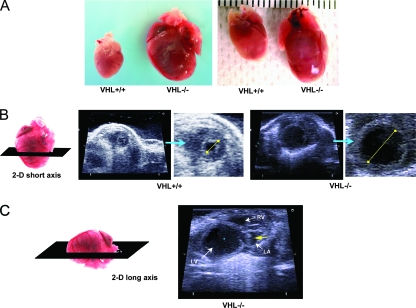
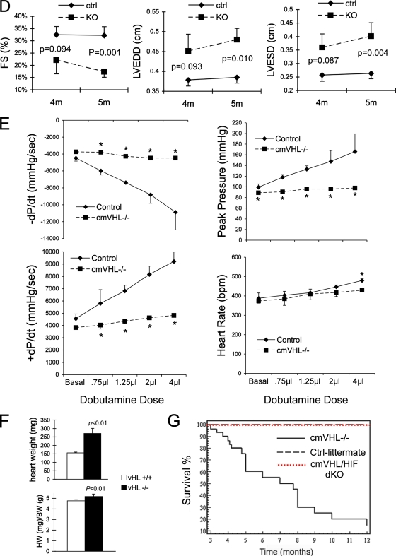
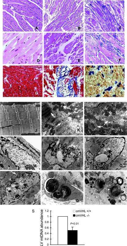
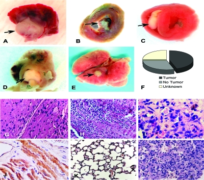
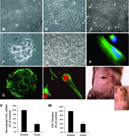
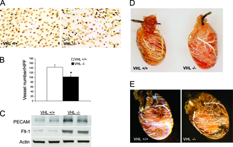
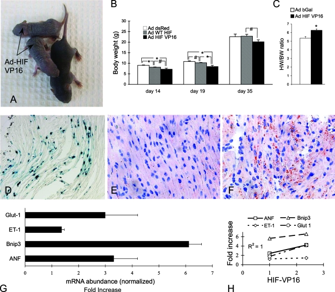
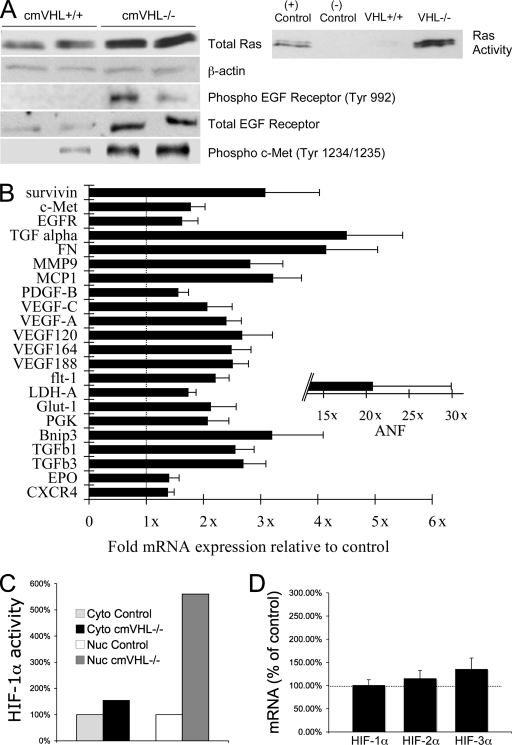
Hypoxia-inducible transcription factor 1 (HIF-1) and HIF-2alpha regulate the expression of an expansive array of genes associated with cellular responses to hypoxia. Although HIF-regulated genes mediate crucial beneficial short-term biological adaptations, we hypothesized that chronic activation of the HIF pathway in cardiac muscle, as occurs in advanced ischemic heart disease, is detrimental. We generated mice with cardiac myocyte-specific deletion of the von Hippel-Lindau protein (VHL), an essential component of an E3 ubiquitin ligase responsible for suppressing HIF levels during normoxia. These mice were born at expected frequency and thrived until after 3 months postbirth, when they developed severe progressive heart failure and premature death. VHL-null hearts developed lipid accumulation, myofibril rarefaction, altered nuclear morphology, myocyte loss, and fibrosis, features seen for various forms of human heart failure. Further, nearly 50% of VHL(-/-) hearts developed malignant cardiac tumors with features of rhabdomyosarcoma and the capacity to metastasize. As compelling evidence for the mechanistic contribution of HIF-1alpha, the concomitant deletion of VHL and HIF-1alpha in the heart prevented this phenotype and restored normal longevity. These findings strongly suggest that chronic activation of the HIF pathway in ischemic hearts is maladaptive and contributes to cardiac degeneration and progression to heart failure.


Figures

FIG. 1.
FIG. 1.
Targeted deletion of VHL in cardiac myocytes leads to cardiomegaly, progressive heart failure, tumor formation, and cardiac death. (A) Cardiac myocyte-specific deletion of VHL (VHL−/−) results in development of severe cardiomegaly relative to what is seen for hearts from age- and sex-matched littermates (VHL+/+). (B) Short-axis two-dimensional (2-D) echocardiography (orientation of ultrasound slice depicted at left) demonstrates severe left ventricular dilation in VHL−/− hearts. For VHL+/+ and VHL−/− hearts, the image on the right is an enlargement of the short-axis image. The yellow lines depict relative intraventricular diameters during diastole. (C) Long-axis two-dimensional echocardiography (orientation depicted at left) demonstrating chamber dilation and the presence of a tumor in the left atrium. LV, left ventricle; RV, right ventricle; LA, left atrium; the yellow arrow depicts a tumor. (D) Echocardiographic analysis of cardiac function reveals significantly reduced fractional shortening during contraction of VHL−/− hearts and significant increases in both end-diastolic and end-systolic diameters of the left ventricle. Whereas these differences were significant at 5 months postbirth, these differences were not significant at 4 months, thus demonstrating the temporal progression of dysfunction and dilation. n ≥ 10 mice per genotype. FS, fractional shortening; LVEDD, left ventricular end-diastolic diameter; LVESD, left ventricular end-systolic diameter; 4m and 5m, 4 and 5 months postbirth; ctrl, control littermates; KO, VHL−/−. (E) Hemodynamic assessment reveals reduced rates of pressure increase and pressure decrease during left ventricular contraction in VHL−/− hearts (+dP/dt and −dP/dt, respectively) and reduced peak developed pressures in VHL−/− hearts at baseline and during progressive infusion rates of dobutamine. Heart rates were similar in VHL−/− and VHL+/+ (control littermate) hearts at all but the highest dose of dobutamine. (F) Heart weights and heart weight/body weight ratios were higher for the cmVHL−/− mice (n ≥ 12 per genotype). (G) Cardiac deletion of VHL results in early and progressive mortality, beginning after 3 months postbirth. Concomitant deletion of VHL and HIF-1α in heart muscle prevents this increased mortality. dKO, double knockout. Curve generated with ≥20 mice per genotype.
FIG. 1.
FIG. 1.
Targeted deletion of VHL in cardiac myocytes leads to cardiomegaly, progressive heart failure, tumor formation, and cardiac death. (A) Cardiac myocyte-specific deletion of VHL (VHL−/−) results in development of severe cardiomegaly relative to what is seen for hearts from age- and sex-matched littermates (VHL+/+). (B) Short-axis two-dimensional (2-D) echocardiography (orientation of ultrasound slice depicted at left) demonstrates severe left ventricular dilation in VHL−/− hearts. For VHL+/+ and VHL−/− hearts, the image on the right is an enlargement of the short-axis image. The yellow lines depict relative intraventricular diameters during diastole. (C) Long-axis two-dimensional echocardiography (orientation depicted at left) demonstrating chamber dilation and the presence of a tumor in the left atrium. LV, left ventricle; RV, right ventricle; LA, left atrium; the yellow arrow depicts a tumor. (D) Echocardiographic analysis of cardiac function reveals significantly reduced fractional shortening during contraction of VHL−/− hearts and significant increases in both end-diastolic and end-systolic diameters of the left ventricle. Whereas these differences were significant at 5 months postbirth, these differences were not significant at 4 months, thus demonstrating the temporal progression of dysfunction and dilation. n ≥ 10 mice per genotype. FS, fractional shortening; LVEDD, left ventricular end-diastolic diameter; LVESD, left ventricular end-systolic diameter; 4m and 5m, 4 and 5 months postbirth; ctrl, control littermates; KO, VHL−/−. (E) Hemodynamic assessment reveals reduced rates of pressure increase and pressure decrease during left ventricular contraction in VHL−/− hearts (+dP/dt and −dP/dt, respectively) and reduced peak developed pressures in VHL−/− hearts at baseline and during progressive infusion rates of dobutamine. Heart rates were similar in VHL−/− and VHL+/+ (control littermate) hearts at all but the highest dose of dobutamine. (F) Heart weights and heart weight/body weight ratios were higher for the cmVHL−/− mice (n ≥ 12 per genotype). (G) Cardiac deletion of VHL results in early and progressive mortality, beginning after 3 months postbirth. Concomitant deletion of VHL and HIF-1α in heart muscle prevents this increased mortality. dKO, double knockout. Curve generated with ≥20 mice per genotype.
FIG. 2.
FIG. 2.
Absence of VHL from cardiac myocytes results in myocyte disassembly and loss, replacement fibrosis, automacrophagocytosis, lipid accumulation, organelle inclusions, and an altered nuclear envelope. (A to C) Hematoxylin and eosin staining (200× magnification) of myocardia from mice with cardiac myocyte-specific deletion of VHL (cmVHL−/−) reveals severe myocardial degeneration with thinning and loss of cardiac muscle bundles (B and C) compared to VHL+/+ control littermate hearts (A). Also visible is patchy infiltration by inflammatory cells (C). (D to F) Analysis at 400× magnification further demonstrates myocardial degeneration and nonuniformity of cardiac muscle bundles (E and F) and cellular infiltration (F) compared to control myocardium (D). (G and H) Myocyte loss and replacement fibrosis is also shown by Mason's trichrome staining of cmVHL−/− hearts (H) versus control littermates (G). (I) cmVHL−/− hearts also accumulate lipids, as shown by oil red O staining. (J to L) Ultrastructural analysis by transmission EM demonstrates disarray and disassembly of myofibrils (white arrows), irregular spacing of Z-bands, irregular orientation of myofibrils, and mitochondrial inclusions (yellow arrow) in cmVHL−/− hearts (K and L) versus the normal architecture of control hearts (J). (N to P) Nuclei from cmVHL−/− hearts exhibit irregular nuclear morphology with prominent folding and blebbing of the nuclear envelope (blue arrows) and multiple nuclear inclusions (black arrows) compared to the normal nuclear architecture in control myocardium (M; arrowhead indicates nucleolus). (Q and R) Multilaminar vesicles (autophagosomes) containing whole organelles (e.g., mitochondria), myofibrils, and other cellular debris were seen frequently in cmVHL−/− hearts, indicating increased autophagy/macroautophagy. (S) Quantitative PCR for mitochondrial DNA revealed a decrease in cmVHL−/− hearts (n = 5 per genotype). For ultrastructural and histological analysis, ≥5 hearts per genotype were studied, with at least five sections and five separate fields/section evaluated per heart.
FIG. 3.
FIG. 3.
Cardiac myocyte-specific deletion of VHL results in primary cardiac tumors in the heart that have the capacity to metastasize. (A to D) Spontaneous primary cardiac tumors growing from the atrial-ventricular junction into the left atrium (A and D), the intraventricular septum into the right ventricle (C), and the left ventricular free wall into the left ventricular chamber (D). Areas from which tumors develop are areas in which MLC2V-Cre is expressed and therefore where VHL is excised. (E) Metastatic tumor to lung. (F) Graphic representation of the incidence of tumors in cmVHL−/− mice. (G to I) Hematoxylin and eosin-stained sections of cmVHL−/− myocardium with tumor infiltration. At a magnification of ×200, the loss of myocardial normal myocardial architecture (J) (compare with Fig. 2A) and myocardial replacement by undifferentiated cells (G and H) are seen. (I) At a magnification of ×400, variable morphologies of nuclei and cells are seen. (J) Tumor tissue exhibits desmin staining and histological characteristics of myofibrillar structure, features consistent with rhabdomyosarcoma. (K) Normal lung histology from control littermate. (L) Histology of metastatic tumor in lung tissue. (M to U) Cultured tumor cells from cmVHL−/− hearts display multiple morphologies (M and N), lack contact inhibition (O), form colonies in soft agarose (P), and can form myotubes (Q). These cells can also become multinucleate and form myofibril-like structures (R), are desmin positive (S and T) (desmin staining is seen as green fluorescence), and form tumors in immunodeficient mice when injected subcutaneously (U) (arrow depicts tumor; n = 4). Quantitative RT-PCR (V) and quantitative PCR (W) of tumors revealed reduced VHL expression correlating with VHL gene excision.
FIG. 3.
FIG. 3.
Cardiac myocyte-specific deletion of VHL results in primary cardiac tumors in the heart that have the capacity to metastasize. (A to D) Spontaneous primary cardiac tumors growing from the atrial-ventricular junction into the left atrium (A and D), the intraventricular septum into the right ventricle (C), and the left ventricular free wall into the left ventricular chamber (D). Areas from which tumors develop are areas in which MLC2V-Cre is expressed and therefore where VHL is excised. (E) Metastatic tumor to lung. (F) Graphic representation of the incidence of tumors in cmVHL−/− mice. (G to I) Hematoxylin and eosin-stained sections of cmVHL−/− myocardium with tumor infiltration. At a magnification of ×200, the loss of myocardial normal myocardial architecture (J) (compare with Fig. 2A) and myocardial replacement by undifferentiated cells (G and H) are seen. (I) At a magnification of ×400, variable morphologies of nuclei and cells are seen. (J) Tumor tissue exhibits desmin staining and histological characteristics of myofibrillar structure, features consistent with rhabdomyosarcoma. (K) Normal lung histology from control littermate. (L) Histology of metastatic tumor in lung tissue. (M to U) Cultured tumor cells from cmVHL−/− hearts display multiple morphologies (M and N), lack contact inhibition (O), form colonies in soft agarose (P), and can form myotubes (Q). These cells can also become multinucleate and form myofibril-like structures (R), are desmin positive (S and T) (desmin staining is seen as green fluorescence), and form tumors in immunodeficient mice when injected subcutaneously (U) (arrow depicts tumor; n = 4). Quantitative RT-PCR (V) and quantitative PCR (W) of tumors revealed reduced VHL expression correlating with VHL gene excision.
FIG. 4.
FIG. 4.
cmVHL−/− hearts exhibit nonuniform hypovascularity. (A and B) Anti-PECAM immunostaining reveals a significant decrease in average capillary counts in cmVHL−/− hearts. (C) Despite decreased average capillary counts, there was an increase in total PECAM and Flt-1 protein in cmVHL−/− hearts, possibly attributed to myocardial infiltration by PECAM/Flt-1-positive inflammatory cells (Fig. 2C and F). (D and E) Casts of the coronary vasculature demonstrate regional variability and decreased macrovascular density in cmVHL−/− hearts. *, n ≥ 5 hearts per genotype for vessel counts; n = 4 hearts per genotype for casts.
FIG. 5.
FIG. 5.
Forced cardiac overexpression of HIF-1α results in myocardial lipid accumulation and failure to thrive. On day 1 postbirth, adenovirus encoding either beta-galactosidase (Ad-β-Gal [Ad bGal]) or a stable form of HIF-1α (Ad-HIFVP16) was delivered to the hearts of neonatal mice. (A to C) Neonatal expression of Ad-HIFVP16 in the heart resulted in significant growth retardation and increased heart weight/body weight ratios 10 days postbirth (#, P = 0.07; *, P < 0.05). WT, wild type. (D) Illustration of the general efficiency of gene delivery to the heart 10 days after neonatal Ad-β-Gal gene delivery. (E and F) Oil red O staining reveals a marked increase in myocardial lipid content in Ad-HIFVP16 hearts (F) versus those that received Ad-β-Gal (E). (G) Ad-HIFVP16 expression in the neonatal heart results in marked induction of HIF-responsive genes (assessment by real-time RT-PCR; values relative to those for Ad-β-Gal hearts and normalized to 18S; onefold is baseline expression). (H) Induction of gene expression correlates with the efficiency of gene delivery as defined by HIF-VP16 expression in the heart (basal VP16 value, 0; VP16 of 1 was the lowest level detected). ANF, atrial natriuretic factor; ET-1, endothelin 1; Bnip3, bcl2/adenovirus E1B-interacting protein 3; Glut-1, glucose transporter 1. n ≥ 5 per group.
FIG. 6.
FIG. 6.
VHL deletion in cardiac myocytes is associated with Ras activation, EGFR and cMET phosphorylation, altered expression of HIF-responsive genes, and increased HIF-1α HRE binding activity. (A) cmVHL−/− hearts have higher total and activated Ras levels and higher levels of phosphorylated (active) cMET and EGFR. (B) Real-time RT-PCR revealed significant alterations in the expression of a variety of HIF-responsive genes (normalized for 18S; “1x” indicates identical expression with control littermates). FN, fibronectin; MMP9, metalloproteinase 9 (gelatinase); MCP-1, monocyte chemotactic protein 1; PDGF, platelet-derived growth factor B; PGK, phosphoglycerate kinase; Glut1, glucose transporter 1; Bnip3, bcl2/adenovirus E1B-interacting protein; EPO, erythropoietin. (C) Pooled nuclear extracts from cmVHL−/− hearts demonstrate higher HIF-1-specific HRE binding (HIF-1α transcription factor binding ELISA). Cyto, cytoplasmic extract; Nuc, nuclear extract. (D) Real-time RT-PCR reveals no significant alterations in mRNA levels for HIF-1 to -3α in cmVHL−/− hearts, consistent with VHL posttranslational control of HIF protein levels (mRNA abundance in control littermate hearts depicted as 100% [dotted line]). n ≥ 5 hearts per genotype.

Similar articles

Cited by

References

    1. Altieri, D. C. 2008. Survivin, cancer networks and pathway-directed drug discovery. Nat. Rev. 861-70. - PubMed
    1. Armstrong, M. T., D. Y. Lee, and P. B. Armstrong. 2000. Regulation of proliferation of the fetal myocardium. Dev. Dyn. 219226-236. - PubMed
    1. Bishop, T., K. W. Lau, A. C. Epstein, S. K. Kim, M. Jiang, D. O'Rourke, C. W. Pugh, J. M. Gleadle, M. S. Taylor, J. Hodgkin, and P. J. Ratcliffe. 2004. Genetic analysis of pathways regulated by the von Hippel-Lindau tumor suppressor in Caenorhabditis elegans. PLoS Biol. 2e289. - PMC - PubMed
    1. Burke, B., and C. L. Stewart. 2002. Life at the edge: the nuclear envelope and human disease. Nat. Rev. 3575-585. - PubMed
    1. Chen, J., S. W. Kubalak, and K. R. Chien. 1998. Ventricular muscle-restricted targeting of the RXRalpha gene reveals a non-cell-autonomous requirement in cardiac chamber morphogenesis. Development (Cambridge) 1251943-1949. - PubMed

Publication types

MeSH terms