. 2008 May;76(5):2063-9.
doi: 10.1128/IAI.01643-07. Epub 2008 Feb 19.

LipL32 is an extracellular matrix-interacting protein of Leptospira spp. and Pseudoalteromonas tunicata

Affiliations

LipL32 is an extracellular matrix-interacting protein of Leptospira spp. and Pseudoalteromonas tunicata

David E Hoke et al. Infect Immun. 2008 May.

Abstract

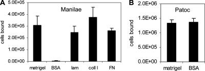
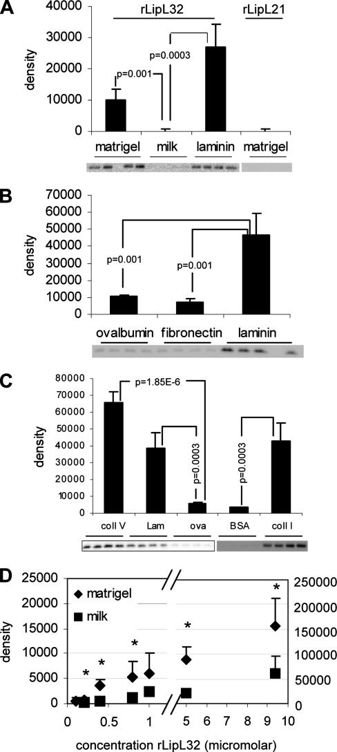
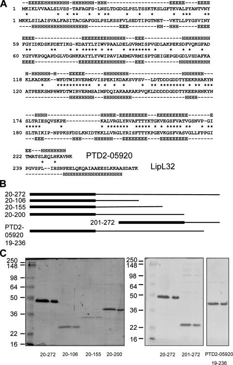
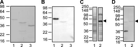
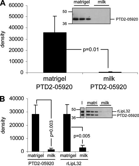
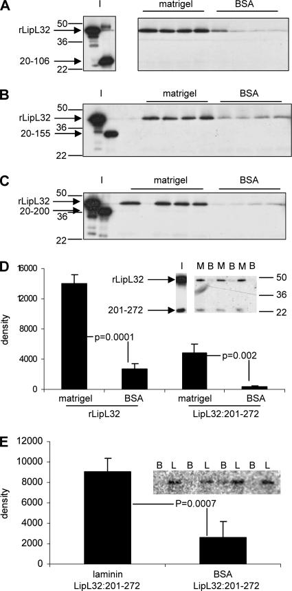
LipL32 is the major outer membrane protein in pathogenic Leptospira. It is highly conserved throughout pathogenic species and is expressed in vivo during human infection. While these data suggest a role in pathogenesis, a function for LipL32 has not been defined. Outer membrane proteins of gram-negative bacteria are the first line of molecular interaction with the host, and many have been shown to bind host extracellular matrix (ECM). A search for leptospiral ECM-interacting proteins identified the major outer membrane protein, LipL32. To verify this finding, recombinant LipL32 was expressed in Escherichia coli and was found to bind Matrigel ECM and individual components of ECM, including laminin, collagen I, and collagen V. Likewise, an orthologous protein found in the genome of Pseudoalteromonas tunicata strain D2 was expressed and found to be functionally similar and immunologically cross-reactive. Lastly, binding activity was mapped to the C-terminal 72 amino acids. These studies show that LipL32 and an orthologous protein in P. tunicata are immunologically cross-reactive and function as ECM-interacting proteins via a conserved C-terminal region.


Figures

FIG. 1.
FIG. 1.
L. interrogans serovar Manilae specifically adheres to surfaces coated with ECM components, while L. biflexa serovar Patoc binds nonspecifically. (A) Wells were coated with Matrigel, BSA, laminin (lam), collagen I (coll I), or fibronectin (FN). The values shown are the averages and standard deviations of the numbers of L. interrogans serovar Manilae cells bound from three replicate wells. Matrigel, laminin, collagen I, and fibronectin binding data were compared to BSA by Student's two-tailed t test with the resulting P values being <0.004. (B) Binding levels of L. biflexa serovar Patoc to Matrigel and BSA were equivalent.
FIG. 2.
FIG. 2.
rLipL32 specifically binds Matrigel, laminin, collagen I, and collagen V. (A to C) Wells of a 96-well plate were coated in quadruplicate with Matrigel, milk, or laminin (A); ovalbumin, fibronectin, or laminin (B); or collagen V (coll V), laminin (lam), ovalbumin (ova), bovine serum albumin (BSA), or collagen I (coll I) (C). The blots below the graphs show rLipL32 or rLipL21 recovered from each binding substrate. The bands were analyzed by densitometry, with the average and standard deviation of each binding condition shown in the bar graphs as indicated. Conditions were compared by Student's two-tailed t test, with the resulting P values indicated. (D) Concentrations of rLipL32 were added to Matrigel- or BSA-control-treated wells, and the amounts of bound protein were determined as described for panels A to C in triplicate. The asterisks above the data points indicate P values of <0.04 for the amount of rLipL32 bound to Matrigel compared to BSA (control).
FIG. 3.
FIG. 3.
Sequence analysis of LipL32 and an orthologous protein, PTD2-05920, from P. tunicata. (A) Alignment of PTD2-05920 and LipL32 sequences. Identical amino acids are indicated by asterisks. Predicted β-sheet regions are designated by E, while α-helices are denoted by H. A predicted random coil is denoted by a dash. (B) rLipL32 deletion derivatives and PTD2-05920 proteins used in this study. The N-terminal biotin acceptor peptide tag is denoted by the thick lines at the left, while LipL32 or PTD2-05920 sequence is shown as a thin line to the right (drawn to scale). The amino acid numbers comprising each construct are shown on the left. (C) Purification of LipL32 deletion derivatives and PTD2-05920. The positions of standard molecular mass markers (kDa) are shown on the left. Purified proteins were resolved in duplicate lanes as indicated: rLipL32, 20 to 272; rLipL32 deletion, 20 to 106; rLipL32 deletion, 20 to 155; rLipL32 deletion, 20 to 200; rLipL32 deletion, 201 to 272; and recombinant PTD2-05920, 19 to 236.
FIG. 4.
FIG. 4.
rPTD2-05920 is recognized by anti-LipL32 serum. (A) rLipL32 (lane 1), rPTD2-05920 (lane 2), and rLipL21 (lane 3) were analyzed by Coomassie blue staining of SDS-PAGE-separated proteins. The positions of standard molecular mass markers (kDa) are shown on the left. rPTD2-05920 migrates at a lower apparent molecular mass than rLipL32 due to the fact that it is 36 amino acids smaller. (B) The same proteins as in panel A were analyzed with anti-LipL32 antiserum by Western blotting. (C) The lysate from 108 L. interrogans cells (lane 1) and the lysate from P. tunicata strain D2 (14 μg) (lane 2) were analyzed by Coomassie blue staining of SDS-PAGE-separated proteins. A highly abundant protein in P. tunicata is indicated by the arrowhead. (D) The same proteins as in panel C were probed with anti-LipL32 antiserum, showing reactivity with LipL32 (lane 1) but not with PTD2-05920. Nonspecific binding to the prominent P. tunicata band (identified as PTD2-07619) (see Results) is indicated by the arrowhead.
FIG. 5.
FIG. 5.
rPTD2-05920 binds Matrigel. (A) rPTD2-05920 was added to Matrigel- or milk-treated wells in triplicate. The blot shows rPTD2-05920 recovered from each binding substrate. The positions of standard molecular mass markers (kDa) are shown on the left. The bands were analyzed by densitometry, with the averages and standard deviations shown in the bar graph. Conditions were compared by Student's two-tailed t test, with the resulting P values indicated. (B) Equivalent amounts of rLipL32 and rPTD2-05920 were incubated with Matrigel (matri) or milk wells and analyzed as described for panel A. The input ratio of rLipL32 and rPTD2-05920 is shown in lane I.
FIG. 6.
FIG. 6.
The C-terminal 72 amino acids of LipL32 are necessary and sufficient for binding to Matrigel and laminin. (A) rLipL32 was coincubated with rLipL32 deletion 20 to 106 at the input ratio shown in blot I. This mixture was incubated with Matrigel- or BSA-treated wells in quadruplicate. The blot shows that only rLipL32 was recovered from the Matrigel-treated wells. The positions of standard molecular mass markers (kDa) are shown on the left. (B) rLipL32 deletion 20 to 155 was coincubated with rLipL32 at the input ratio shown in lanes I. Proteins were added to Matrigel- or BSA-treated wells as for panel A. Only rLipL32 was recovered from Matrigel-treated wells. (C) rLipL32 deletion 20 to 200 was coincubated with rLipL32 and analyzed for binding as for panel A. Only rLipL32 was recovered in the Matrigel-treated wells. (D) rLipL32 deletion 201 to 272 was coincubated with rLipL32 at the input ratio indicated by lane I. Both proteins were recovered from Matrigel-treated wells (denoted by M) at similar ratios. Neither protein was recovered from BSA-treated wells (denoted by B). The positions of standard molecular mass markers are shown on the right. The blot was quantified by densitometry, with the averages and standard deviations from triplicate wells presented graphically. Conditions were compared by Student's two-tailed t test, with the resulting P values indicated. (E) rLipL32 deletion 201 to 272 was incubated with laminin (L) or BSA control (B) in quadruplicate. The blot shows the amount of rLipL32 deletion 201 to 272 recovered. The bands were analyzed by densitometry as in panel D.

References

    1. Anonymous. 1999. Leptospirosis worldwide, 1999. Wkly. Epidemiol. Rec. 74217-223. - PubMed
    1. Barbosa, A. S., P. A. Abreu, F. O. Neves, M. V. Atzingen, M. M. Watanabe, M. L. Vieira, Z. M. Morais, S. A. Vasconcellos, and A. L. Nascimento. 2006. A newly identified leptospiral adhesin mediates attachment to laminin. Infect. Immun. 746356-6364. - PMC - PubMed
    1. Choy, H. A., M. M. Kelley, T. L. Chen, A. K. Moller, J. Matsunaga, and D. A. Haake. 2007. Physiological osmotic induction of Leptospira interrogans adhesion: LigA and LigB bind extracellular matrix proteins and fibrinogen. Infect. Immun. 752441-2450. - PMC - PubMed
    1. Cullen, P. A., S. J. Cordwell, D. M. Bulach, D. A. Haake, and B. Adler. 2002. Global analysis of outer membrane proteins from Leptospira interrogans serovar Lai. Infect. Immun. 702311-2318. - PMC - PubMed
    1. Cullen, P. A., D. A. Haake, and B. Adler. 2004. Outer membrane proteins of pathogenic spirochetes. FEMS Microbiol. Rev. 28291-318. - PMC - PubMed

Publication types

MeSH terms

LinkOut - more resources