. 2008 Feb 20;3(2):e1614.
doi: 10.1371/journal.pone.0001614.

A non-human primate model for gluten sensitivity

Affiliations

A non-human primate model for gluten sensitivity

Michael T Bethune et al. PLoS One. .

Abstract

Background and aims: Gluten sensitivity is widespread among humans. For example, in celiac disease patients, an inflammatory response to dietary gluten leads to enteropathy, malabsorption, circulating antibodies against gluten and transglutaminase 2, and clinical symptoms such as diarrhea. There is a growing need in fundamental and translational research for animal models that exhibit aspects of human gluten sensitivity.

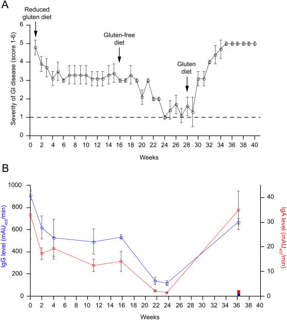
Methods: Using ELISA-based antibody assays, we screened a population of captive rhesus macaques with chronic diarrhea of non-infectious origin to estimate the incidence of gluten sensitivity. A selected animal with elevated anti-gliadin antibodies and a matched control were extensively studied through alternating periods of gluten-free diet and gluten challenge. Blinded clinical and histological evaluations were conducted to seek evidence for gluten sensitivity.

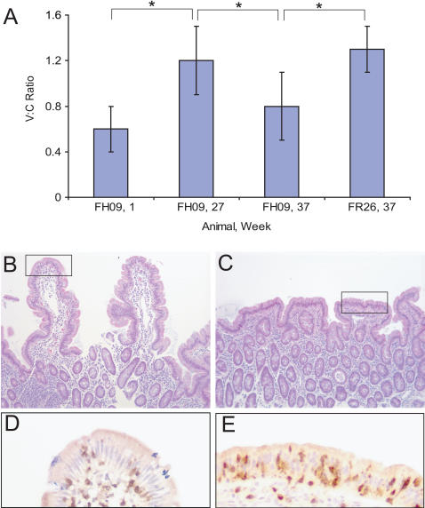
Results: When fed with a gluten-containing diet, gluten-sensitive macaques showed signs and symptoms of celiac disease including chronic diarrhea, malabsorptive steatorrhea, intestinal lesions and anti-gliadin antibodies. A gluten-free diet reversed these clinical, histological and serological features, while reintroduction of dietary gluten caused rapid relapse.

Conclusions: Gluten-sensitive rhesus macaques may be an attractive resource for investigating both the pathogenesis and the treatment of celiac disease.


Conflict of interest statement

Competing Interests: The authors have declared that no competing interests exist.

Figures

Figure 1
Figure 1. Histopathology of the small intestine.
H&E-stained tissue sections of duodenum and proximal jejunum from rhesus macaques with idiopathic diarrhea. (A) Normal control duodenum from an age-matched rhesus macaque illustrating characteristic morphology of the villi. 100× magnification. (B) Enteropathy of duodenum. Diffuse enteritis characterized by shortening of villi, severe lymphocytic and plasmacytic infiltration of the lamina propria, and vacuolar degeneration of the epithelium. 100× magnification. (C) Normal control jejunum from an age-matched rhesus macaque. 100× magnification. (D) Enteropathy of jejunum. The mucosa appears flat with marked blunting of villi and dense infiltration of lamina propria by mononuclear cells. 100× magnification.
Figure 2
Figure 2. Gluten dependence of clinical symptoms and serology in gluten-sensitive rhesus macaque FH09.
(A) Gastrointestinal symptoms in gluten-sensitive FH09 improve with sequential administration of reduced gluten and gluten-free diets, but return upon reintroduction of dietary gluten (diet changes indicated by vertical arrows). Criteria that were used in clinical scoring of gluten sensitivity in the juvenile macaque FH09 were scaled relative to the healthy, age-matched control, FR26 (score 1, indicated by dotted line). Score 2 corresponded to beginning of diarrhea, e.g. pasty stools. Score 3 corresponded to semi-liquid stools and decreased activity. Score 4 corresponded to liquid stools, decreased activity, moderate dehydration and “balloon” stomach. Score 5 corresponded to liquid stools, depression, severe dehydration and balloon stomach. Score 6 would correspond to a moribund animal where prompt euthanasia is recommended. Each datapoint represents the mean of 7 daily measurements taken over the course of the indicated week. Standard deviations are indicated by error bars. (B) Anti-gliadin IgG (blue; open circles) and IgA (red; open squares) return to baseline with dietary exclusion of gluten, but are elevated following reintroduction of dietary gluten. The level of anti-gliadin IgG (blue; closed circle) and IgA (red; closed square) in control FR26 are shown for comparison. Each datapoint represents the mean of triplicate measurements. Standard deviations are indicated by error bars.
Figure 3
Figure 3. Gluten dependence of histological lesions in gluten-sensitive rhesus macaque FH09.
(A) Morphometric analysis of villus height:crypt depth ratios from at least 4 different areas of distal duodenum in gluten-sensitive FH09 and control FR26 following dietary changes. Administration of a gluten-free diet increased the V:C ratio in FH09 at week 27 to a level that is statistically equivalent to that in FR26 (constant at all time points). Reintroduction of dietary gluten resulted in a drop in V:C ratio in FH09 (week 37) relative to that in FH09 on a gluten-free diet (week 27) and to that in FR26. *P<0.05. (B–C) H&E-stained duodenum at week 37 following 10 weeks of a gluten-containing diet. 100× magnification. (B) Control macaque FR26 exhibits normal villus architecture. (C) Gluten-sensitive macaque FH09 exhibits villus blunting. (D–E) Highlighted sections in B–C were examined by immunohistochemistry. 400× magnification. (D) Anti-CD3 staining in FR26 shows few CD3+ IELs (dark brown dots in epithelium). (E) Anti-CD3 staining in FH09 shows intraepithelial lymphocytosis.

References

    1. Dicke WK, Weijers HA, Van De Kamer JH. Coeliac disease. II. The presence in wheat of a factor having a deleterious effect in cases of coeliac disease. Acta Paediatr. 1953;42:34–42. - PubMed
    1. Alaedini A, Green PH. Narrative review: celiac disease: understanding a complex autoimmune disorder. Ann Intern Med. 2005;142:289–298. - PubMed
    1. Green PH, Jabri B. Celiac disease. Annu Rev Med. 2006;57:207–221. - PubMed
    1. Sollid LM, Markussen G, Ek J, Gjerde H, Vartdal F, et al. Evidence for a primary association of celiac disease to a particular HLA-DQ alpha/beta heterodimer. J Exp Med. 1989;169:345–350. - PMC - PubMed
    1. Spurkland A, Ingvarsson G, Falk ES, Knutsen I, Sollid LM, et al. Dermatitis herpetiformis and celiac disease are both primarily associated with the HLA-DQ (alpha 1*0501, beta 1*02) or the HLA-DQ (alpha 1*03, beta 1*0302) heterodimers. Tissue Antigens. 1997;49:29–34. - PubMed

Publication types