. 2008 Feb 20;3(2):e1624.
doi: 10.1371/journal.pone.0001624.

Isolation, cloning and structural characterisation of boophilin, a multifunctional Kunitz-type proteinase inhibitor from the cattle tick

Affiliations

Isolation, cloning and structural characterisation of boophilin, a multifunctional Kunitz-type proteinase inhibitor from the cattle tick

Sandra Macedo-Ribeiro et al. PLoS One. .

Abstract

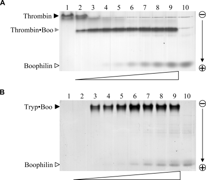
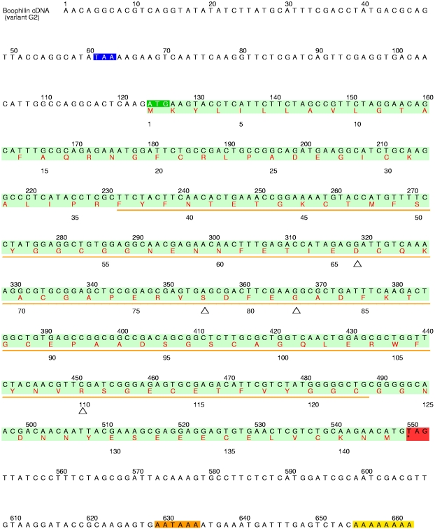
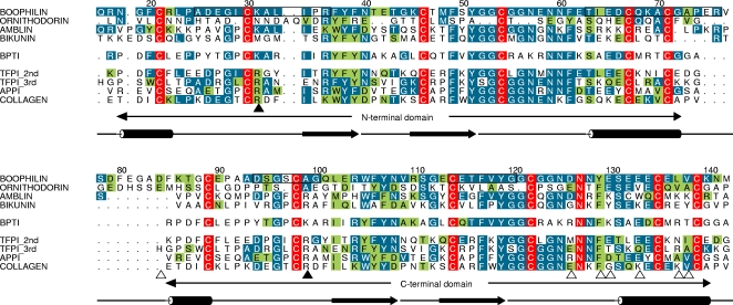
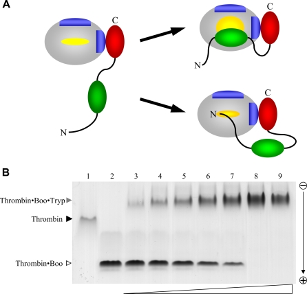
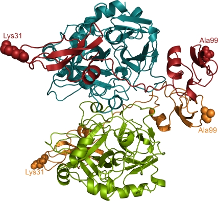
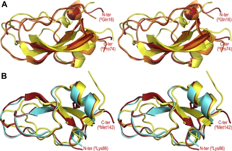
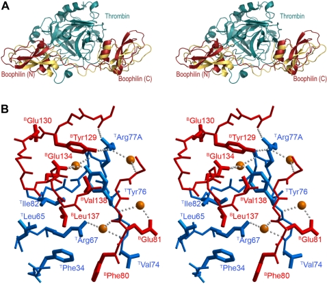
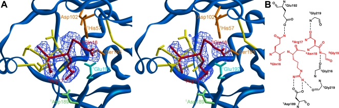
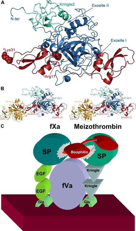
Inhibitors of coagulation factors from blood-feeding animals display a wide variety of structural motifs and inhibition mechanisms. We have isolated a novel inhibitor from the cattle tick Boophilus microplus, one of the most widespread parasites of farm animals. The inhibitor, which we have termed boophilin, has been cloned and overexpressed in Escherichia coli. Mature boophilin is composed of two canonical Kunitz-type domains, and inhibits not only the major procoagulant enzyme, thrombin, but in addition, and by contrast to all other previously characterised natural thrombin inhibitors, significantly interferes with the proteolytic activity of other serine proteinases such as trypsin and plasmin. The crystal structure of the bovine alpha-thrombin.boophilin complex, refined at 2.35 A resolution reveals a non-canonical binding mode to the proteinase. The N-terminal region of the mature inhibitor, Q16-R17-N18, binds in a parallel manner across the active site of the proteinase, with the guanidinium group of R17 anchored in the S(1) pocket, while the C-terminal Kunitz domain is negatively charged and docks into the basic exosite I of thrombin. This binding mode resembles the previously characterised thrombin inhibitor, ornithodorin which, unlike boophilin, is composed of two distorted Kunitz modules. Unexpectedly, both boophilin domains adopt markedly different orientations when compared to those of ornithodorin, in its complex with thrombin. The N-terminal boophilin domain rotates 9 degrees and is displaced by 6 A, while the C-terminal domain rotates almost 6 degrees accompanied by a 3 A displacement. The reactive-site loop of the N-terminal Kunitz domain of boophilin with its P(1) residue, K31, is fully solvent exposed and could thus bind a second trypsin-like proteinase without sterical restraints. This finding explains the formation of a ternary thrombin.boophilin.trypsin complex, and suggests a mechanism for prothrombinase inhibition in vivo.


Conflict of interest statement

Competing Interests: The authors have declared that no competing interests exist.

Figures

Figure 1
Figure 1. Boophilin forms stable stoichiometric complexes with free thrombin and trypsin.
(A) Five-hundred nanograms of free human α-thrombin were incubated with increasing amounts of the purified inhibitor (from ≈140 ng, lane 2, to ≈2.1 µg, lane 9), and samples were resolved in a 10% polyacrylamide gel. Lanes 1 and 10 contain 500 ng thrombin and 2.1 µg boophilin, respectively. Notice formation of a single species corresponding to the 1:1 thrombin·boophilin complex (Thrombin·Boo). (B) Detection of equimolar trypsin·boophilin complex. Five-hundred nanograms bovine trypsin were mixed with increasing amounts of purified boophilin (from ≈100 ng, lane 2, to 1.2 µg, lane 9), and samples were separated in a 10% polyacrylamide gel. Lanes 1 and 10 contain 500 ng trypsin and 1.2 µg boophilin, respectively; cationic trypsin does not migrate into the gel. The stoichiometric trypsin·boophilin complex is marked (Tryp·Boo).
Figure 2
Figure 2. Complete nucleotide sequence of the cDNA encoding for boophilin variant G2 and predicted amino acid sequence.
The coding region is shaded in light green, the start codon in dark green, the stop codon in red, the polyadenylation signal in orange, and the short poly(A) tail in yellow. The deduced amino acid sequence of pro-boophilin is shown in red below the nucleotide sequence, in one-letter code. An orange line indicates the fragment amplified by PCR. The in-frame stop codon that precedes the start codon for pro-boophilin is shaded blue. The deduced amino acid sequence of boophilin (142 residues) would correspond to a polypeptide with a higher molecular mass than that experimentally determined by MALDI-MS. However, if one considers the presence of a 15-residue pro-peptide, the resulting mature inhibitor would have a theoretical mass of 13,950 Da, in reasonable agreement with the experimentally determined value of 13,964 Da. (Notice that no putative N-glycosylation sites are present in the boophilin sequence). Further, the mature protein starts with a glutamine residue that could spontaneously cyclise to pyrrolidone carboxylic acid (5-oxo-proline). This modification would explain the blocked N-terminus observed in native boophilin, and has previously been reported in other Kunitz inhibitors such as an BPTI variant from bovine lung . Open triangles indicate the positions where discrete differences exist in the amino acid sequences of the two boophilin variants.
Figure 3
Figure 3. The two boophilin domains are more closely related to canonical Kunitz modules than to ornithodorin.
Comparison of the amino acid sequence of mature boophilin with three other two-domain inhibitors, ornithodorin, amblin, and bikunin; the unique C-terminal extension in amblin has been omitted for clarity. Also included in the alignment are the sequences of BPTI and of the closest related human Kunitz modules structurally characterised so far (two copies, one aligned with each Kunitz module of boophilin): The second and third TFPI domains (1TFX and 1IRH , respectively), the single Kunitz domain of APP (1AAP) , and the C-terminal Kunitz domain of type VI collagen (2KNT) . Numbers refer to the full-length sequence of boophilin. Residues conserved throughout are white with red shading; residues identical to boophilin are white with blue shading; conservatively replaced residues are shaded green. The four major antigenic determinants in boophilin are boxed. The secondary structure elements of boophilin are shown below the alignment; β-strands are represented as arrows and α-helices as cylinders. The P1 residues within the reactive-site loops are indicated with black arrowheads; open arrowheads point to boophilin/ornithodorin residues involved in important contacts with thrombin exosite I. Notice that several exosite-interacting residues are conserved or conservatively replaced in boophilin, while they are mostly non-conservatively replaced in amblin (e.g., E130→K, L137→K).
Figure 4
Figure 4. Thrombin-bound boophilin retains the ability to interact with other serine proteinases.
(A) Schematic representation of two hypothetical thrombin·boophilin complexes. In the upper, BPTI-like mechanism, binding of the C-terminal boophilin domain to exosite I promotes extensive rearrangements of loops surrounding the active site to allow insertion of the N-terminal domain in a canonical manner. In the alternative, ornithodorin-like mechanism, exosite engagement is not associated with important modifications of the thrombin active site region, which is occupied by the N-terminal peptide of the inhibitor in a parallel manner. (B) Demonstration of thrombin·boophilin·trypsin ternary complex formation via native gel electrophoresis. One µg human α-thrombin·boophilin complex was incubated with increasing amounts of bovine trypsin (from ≈300 ng, lane 3, to ≈6 µg, lane 9), and samples were resolved in an 8% polyacrylamide gel. Lanes 1 and 2 contain 1 µg thrombin and 1 µg thrombin·boophilin complex, respectively; the newly formed species corresponds to the ternary complex.
Figure 5
Figure 5. Three-dimensional structure of the α-thrombin·boophilin complex.
The crystallographic dimer present in the asymmetric unit is shown as a ribbon plot; thrombin molecules are coloured blue and green and boophilin molecules are coloured red and orange. Notice that the reactive-site loops of both inhibitor domains point away from the proteinase moiety; the corresponding P1 residues, BK31 and BA99, are shown as space-filling models. Within the dimer interface, direct hydrogen bonds are formed between the side chain of BS97 and the main-chain carbonyl of BG124 located in opposite C-terminal domains of boophilin, and between the side chains of TD21 and TQ14A.
Figure 6
Figure 6. Boophilin is structurally more related to canonical Kunitz modules than to the distorted ornithodorin domains.
Stereo plots showing the three-dimensional structure of (A) boophilin's N-terminal domain (red) overlaid on those of the second TFPI domain, as seen in its complex with porcine trypsin (orange, PDB entry 1TFX) , and of the N-terminal ornithodorin domain (yellow, 1TOC) , and (B) boophilin's C-terminal domain (red) superimposed on those of APP single Kunitz domain (cyan, 1TAW) , and the C-terminal ornithodorin domain (yellow, 1TOC) . The N and C termini of boophilin are labelled and its disulfide bonds are shown as sticks. Notice that in spite of significant sequence similarity between the carboxy-terminal domain of boophilin and other Kunitz-type inhibitors (Figure 3), with strict conservation of cysteine spacing, its putative P1 residue is nevertheless an alanine (BA99), thus precluding inhibition of trypsin-like serine proteinases.
Figure 7
Figure 7. Different orientations of the Kunitz domains in thrombin complexes with boophilin and ornithodorin.
(A) Stereo plot showing a comparison of thrombin·boophilin (blue/red) and thrombin·ornithodorin (blue/yellow) complexes, after overlaying both thrombin moieties. Notice the large displacements of the corresponding N- and C-terminal Kunitz modules relative to each other. (B) Stereo close-up highlighting the docking of the C-terminal domain of boophilin (red) to thrombin (blue) exosite I. Water molecules are represented as orange spheres and hydrogen bonds as rows of small grey spheres. The side chains of residues involved in intermolecular contacts are shown and labelled.
Figure 8
Figure 8. Boophilin inhibits thrombin in a non-canonical manner.
(A) Stereo close-up of the thrombin active centre (blue) showing the bound tetrapeptide BQ16–BR17–BN18–BG19 of boophilin (red), along with S1A–L1–N2–V3 of ornithodorin (yellow; notice that the N-terminal residue of the latter is an artifact introduced for cloning purposes). The final electron density for the thrombin·boophilin complex, contoured at 1σ, is displayed as a blue mesh. The catalytic triad of thrombin (TH57, TD102, TS195) is highlighted in orange and the side-chains of TD189 and TE192 are coloured pale green and cyan, respectively. Disulfide bonds are represented as yellow sticks. (B) Schematic representation of the thrombin-boophilin interactions at the enzyme's active site. Inhibitor residues are coloured red and hydrogen bonds are depicted as dashed lines.
Figure 9
Figure 9. Predicted boophilin interactions with meizothrombin and FXa.
(A) Model of the three-dimensional meizothrombin·boophilin complex, generated by overlaying the coordinates of bovine meizothrombin (blue except for the Kringle 2 domain, in green; 1A0H) onto the thrombin moiety of the current complex with boophilin. Residues BR17 and BK31 are shown as space-filling models and labelled. (B) Model of the putative MzT·boophilin·FXa ternary complex. FXa (yellow, except for domain EGF2, in olive; 1FJS) was docked onto the complex displayed in (A) by juxtaposition onto a trypsin·APP complex (1TFX) that had been previously overlaid onto the N-terminal domain of boophilin (red). Absence of any steric clashes shows that formation of such a ternary complex in vivo would be feasible. The N- and C-terminal domains of boophilin (Boophilin (N) and Boophilin (C), respectively), the serine proteinase and Kringle 2 domains of meizothrombin (SP (MzT) and Kringle 2, respectively), and the serine proteinase and EGF-2 domains of factor Xa (SP (FXa) and EGF-2, respectively) are labelled. (C) Schematic representation of a possible mechanism for boophilin inhibition of the membrane-bound prothrombinase complex. Both FXa and meizothrombin interact with the membrane surface (dark red) via their respective Gla domains (light green). The multi-domain organisation of FVa (violet) and its contacts with FXa/MzT have been omitted for simplicity. The N-terminal domain of boophilin (red) could bridge the catalytic domains of meizothrombin (dark green) and FXa (blue) while bound to the cofactor.

Similar articles

Cited by

References

    1. Bork P, Downing AK, Kieffer B, Campbell ID. Structure and distribution of modules in extracellular proteins. Q Rev Biophys. 1996;29:119–167. - PubMed
    1. Laskowski M, Jr, Kato I. Protein inhibitors of proteinases. Annu Rev Biochem. 1980;49:593–626. - PubMed
    1. Ascenzi P, Bocedi A, Bolognesi M, Spallarossa A, Coletta M, et al. The bovine basic pancreatic trypsin inhibitor (Kunitz inhibitor): a milestone protein. Curr Protein Pept Sci. 2003;4:231–251. - PubMed
    1. Broze GJ., Jr Tissue factor pathway inhibitor and the revised theory of coagulation. Annu Rev Med. 1995;46:103–112. - PubMed
    1. Huber R, Kukla D, Bode W, Schwager P, Bartels K, et al. Structure of the complex formed by bovine trypsin and bovine pancreatic trypsin inhibitor. II. Crystallographic refinement at 1.9 Å resolution. J Mol Biol. 1974;89:73–101. - PubMed

Publication types