. 2008 Apr 15;56(6):611-8.
doi: 10.1002/glia.20636.

The ABCA1 cholesterol transporter associates with one of two distinct dystrophin-based scaffolds in Schwann cells

Affiliations

The ABCA1 cholesterol transporter associates with one of two distinct dystrophin-based scaffolds in Schwann cells

Douglas E Albrecht et al. Glia. .

Abstract

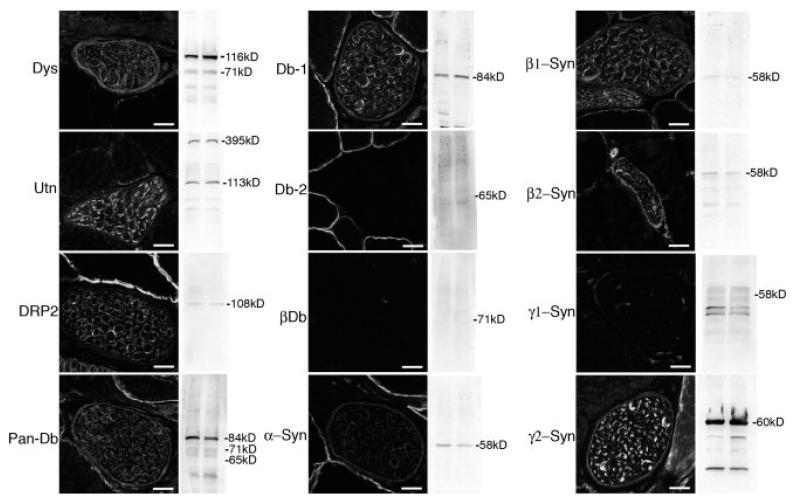
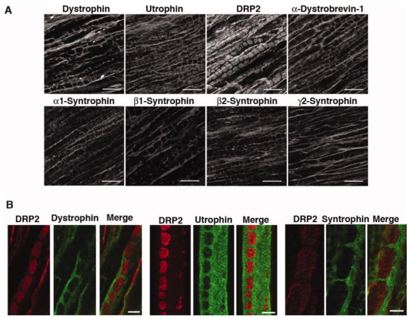
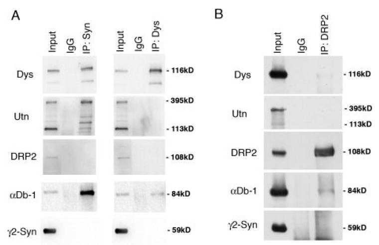
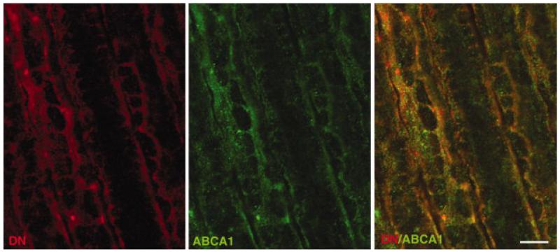
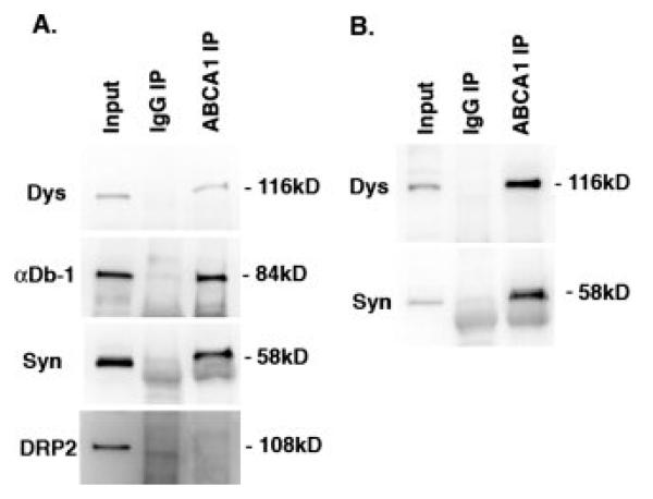
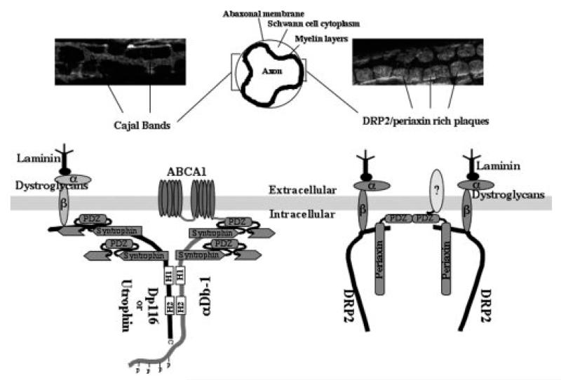
Cytoskeletal scaffolding complexes help organize specialized membrane domains with unique functions on the surface of cells. In this study, we define the scaffolding potential of the Schwann cell dystrophin glycoprotein complex (DGC) by establishing the presence of four syntrophin isoforms, (alpha1, beta1, beta2, and gamma2), and one dystrobrevin isoform, (alpha-dystrobrevin-1), in the abaxonal membrane. Furthermore, we demonstrate the existence of two separate DGCs in Schwann cells that divide the abaxonal membrane into spatially distinct domains, the DRP2/periaxin rich plaques and the Cajal bands that contain Dp116, utrophin, alpha-dystrobrevin-1 and four syntrophin isoforms. Finally, we show that the two different DGCs can scaffold unique accessory molecules in distinct areas of the Schwann cell membrane. Specifically, the cholesterol transporter ABCA1, associates with the Dp116/syntrophin complex in Cajal bands and is excluded from the DRP2/periaxin rich plaques.


Figures

Figure 1
Figure 1. Expression and localization of DGC proteins in peripheral nerve
Immunofluorescence microscopy was used to determine the localization of dystrophin (Dys), utrophin (Utn), DRP2, α-dystrobrevin-1 (Db-1), α-dystrobrevin-2 (Db-2), β-dystrobrevin (βDb), α-, β1, β2, γ1, and γ2-syntrophin (Syn) in nerve branches running through skeletal muscle (transverse sections). Western blots using the corresponding antibody on duplicate samples of whole sciatic nerve homogenates are also shown. Scale bar is 12 μm.
Figure 2
Figure 2
Localization of DGC proteins to Cajal bands and plaques and comparison of the distribution of DRP2 with dystrophin, utrophin, and α/β syntrophin in sciatic nerve. (A) Longitudinal sections of mouse sciatic nerve were stained with the indicated antibodies. Only DRP2 is localized to plaques. Dystrophin, utrophin, α-dystrobrevin-1, and α-, β1-, β2-, and γ2-syntrophin are all restricted to Cajal bands. Scale bar is 10 μm. (B) DRP2 (red) is localized to plaques while dystrophin, utrophin and α/β syntrophin (visualized with mAb 1351) (green) are restricted to Cajal bands. Right and left panels, rat sciatic nerve longitudinal sections. Middle panel, teased fiber. Scale bar is 5 μm.
Figure 3
Figure 3
Coimmunoprecipitation of DGC proteins from mouse sciatic nerve. (A) Detergent extracts (Input) were subjected to immunoprecipitation (IP) with anti-α/β-syntrophin (Syn, mAb 1351), anti-dystrophin (Dys, mandra1), or control antibody (IgG); (B) immunoprecipation with anti-DRP2 or IgG. Immunoprecipitates were subjected to western blotting with the antibodies indicated. Dp116, α/β-syntrophin and α-Db-1 are coimmunoprecipitated. Neither DRP2 nor γ2-syntrophin are present in complexes with Dp116/syn/αDb-1 or with each other.
Figure 4
Figure 4
Colocalization of ABCA1 and Dp116 in Cajal bands. Immunofluorescence labeling of rat sciatic nerve longitudinal sections shows significant, but not complete, colocalization of ABCA1 (green) and Dp116 (DN, Red). Scale bar is 5 μm.
Figure 5
Figure 5
Coimmunoprecipitation of ABCA1 with the Dp116/syn/αDb-1 complex but not with DRP2. (A) Immunoprecipitations from C57 mouse sciatic nerve with ABCA1 were subjected to western blotting for Dp116 (Dys), αDb-1, α/β-syntrophins, and DRP2. (B) A similar experiment using sciatic nerve from the α/β2-syntrophin double null mice. Coimmunoprecipitation of the Dp116/syn/αDb-1 complex with anti-ABCA1 does not depend on the presence of α- or β2-syntrophin. DRP2 is not found in the ABCA1 complex. IgG IP, control.
Figure 6
Figure 6
A model of the dystrophin and DRP2 complexes and their division of the abaxonal membrane into Cajal bands and plaques.

Similar articles

Cited by

References

    1. Adams ME, Butler MH, Dwyer TM, Peters MF, Murnane AA, Froehner SC. Two forms of mouse syntrophin, a 58 kd dystrophin-associated protein, differ in primary structure and tissue distribution. Neuron. 1993;11(3):531–40. - PubMed
    1. Adams ME, Kramarcy N, Fukuda T, Engel AG, Sealock R, Froehner SC. Structural abnormalities at neuromuscular synapses lacking multiple syntrophin isoforms. J Neurosci. 2004;24(46):10302–9. - PMC - PubMed
    1. Adams ME, Kramarcy N, Krall SP, Rossi SG, Rotundo RL, Sealock R, Froehner SC. Absence of alpha-syntrophin leads to structurally aberrant neuromuscular synapses deficient in utrophin. J Cell Biol. 2000;150(6):1385–98. - PMC - PubMed
    1. Adams ME, Mueller HA, Froehner SC. In vivo requirement of the alpha-syntrophin PDZ domain for the sarcolemmal localization of nNOS and aquaporin-4. J Cell Biol. 2001;155(1):113–22. - PMC - PubMed
    1. Albrecht DE, Froehner SC. Syntrophins and dystrobrevins: defining the dystrophin scaffold at synapses. Neurosignals. 2002;11(3):123–9. - PubMed

Publication types

MeSH terms

LinkOut - more resources