Comparative Study
. 2008 Feb 26;105(8):3059-63.
doi: 10.1073/pnas.0712265105. Epub 2008 Feb 14.

Requirement of protein kinase D1 for pathological cardiac remodeling

Affiliations
Comparative Study

Requirement of protein kinase D1 for pathological cardiac remodeling

Jens Fielitz et al. Proc Natl Acad Sci U S A. .

Abstract

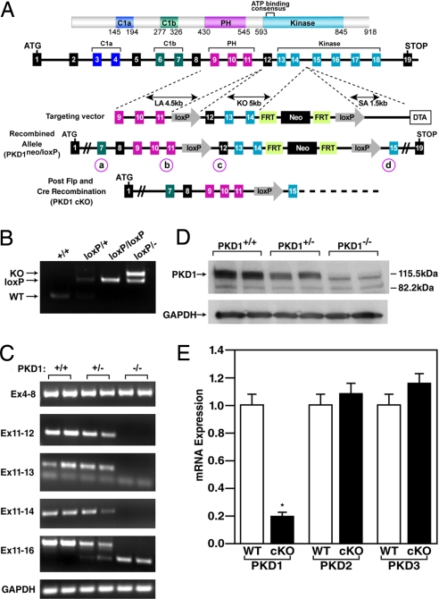
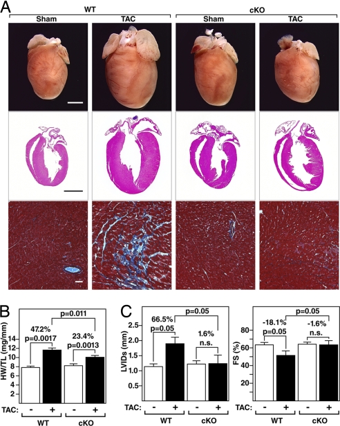
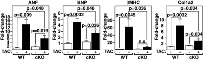
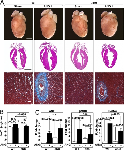
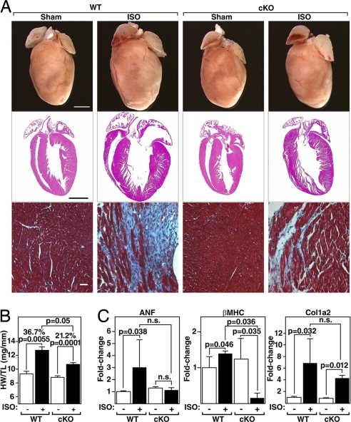
The adult heart responds to biomechanical stress and neurohormonal signaling by hypertrophic growth, accompanied by fibrosis, diminished pump function, and activation of a fetal gene program. Class II histone deacetylases (HDACs) suppress stress-dependent remodeling of the heart via their association with the MEF2 transcription factor, an activator of heart disease. Protein kinase D (PKD) is a stress-responsive kinase that phosphorylates class II HDACs, resulting in their dissociation from MEF2 with consequent activation of MEF2 target genes. To test whether PKD1 is required for pathological cardiac remodeling in vivo, we generated mice with a conditional PKD1-null allele. Mice with cardiac-specific deletion of PKD1 were viable and showed diminished hypertrophy, fibrosis, and fetal gene activation as well as improved cardiac function in response to pressure overload or chronic adrenergic and angiotensin II signaling. We conclude that PKD1 functions as a key transducer of stress stimuli involved in pathological cardiac remodeling in vivo.


Conflict of interest statement

The authors declare no conflict of interest.

Figures

Fig. 1.
Fig. 1.
Generation of mice with a conditional PKD1 mutation. (A) Mouse PKD1 locus and targeting strategy. LoxP sites were inserted in the introns flanking exons 12 and 14. Exons 13 and 14 encode the N-terminal region of the kinase domain including the ATP-binding motif. The neomycin resistance cassette (neo) was removed in the mouse germ line by breeding heterozygous mice to hACTB::FLPe transgenic mice, and deletion of exons 12, 13, and 14 was achieved by breeding PKD1loxP/loxP mice to either CAG-Cre or α-MHC-Cre transgenic mice. Positions of PCR primers used for genotyping are labeled a–d and circled. (B) PCR genotyping to distinguish PKD1 alleles. PCR products corresponding to WT (151 bp), PKD1loxP (loxP) (255 bp), and PKD1 KO (359 bp) are shown. The positions of the primers that produce these PCR products are labeled b and c for WT and PKD1loxP and a and d for PKD1 KO and are circled in A. (C) RT-PCR to detect WT and mutant PKD1 transcripts. The PKD1 mutant allele lacks exons 12, 13, and 14. GAPDH was detected as a loading control. Locations of primers used for RT-PCR are shown on the left. (D) Western blot analysis of PKD1 in cardiac extracts from WT and PKD1 mutant mice. GAPDH protein was used as a loading control. (E) Expression of PKD1 transcripts detected by quantitative PCR. Total RNA isolated from ventricles of 8-week-old male mice was used for cDNA synthesis and subsequent quantitative PCR (n = 6 for each genotype). P < 0.01. Error bars indicate ±SEM.
Fig. 2.
Fig. 2.
Diminished hypertrophy of PKD1 cKO mice after TAC. (A) Hearts from WT and PKD1 mutant mice subjected to either a sham operation (WT and PKD1 cKO, n = 6) or TAC (Top; WT, n = 12; PKD1 cKO, n = 11). Histological sections stained with H&E (Middle) or Masson's trichrome to detect fibrosis (Bottom). (Scale bars: Top and Middle, 2 mm; Bottom, 40 μm.) (B) Heart weight/tibia length (HW/TL) ratios (±SEM) of WT and PKD1 cKO mice were determined 21 days after TAC. (C) PKD1 cKO mice display less left ventricular dilation during systole (LVIDs) and a less pronounced decrease in fractional shortening (FS) in response to TAC than WT mice.
Fig. 3.
Fig. 3.
Diminished fetal gene activation in PKD1 cKO mice after TAC. Transcripts for markers of hypertrophy in hearts from WT and PKD cKO mice were detected by quantitative PCR 21 days after TAC (n = 3–9 per group). Values indicate relative expression level to a WT sham-operated group (±SEM). ANF, atrial natriuretic factor; BNP, brain natriuretic peptide; β-MHC, β-myosin heavy chain; Col1a2, procollagen, type I, α2.
Fig. 4.
Fig. 4.
Diminished hypertrophic response of PKD1 cKO mice to AngII infusion. (A) Hearts of WT and PKD1 cKO mice treated with either saline vehicle (WT and PKD1 cKO, n = 6) or AngII (3.0 mg/kg per day) for 14 days (Top; WT, n = 9; PKD1 cKO, n = 8), histological sections stained with H&E (Middle), or Masson's trichrome to detect fibrosis (Bottom). (Scale bars: Top and Middle, 2 mm; Bottom, 40 μm.) (B) Heart weight/tibia length (HW/TL) ratios (±SEM) are shown as bar graphs (n = 7–9). (C) Transcripts for markers of hypertrophy in hearts from WT and PKD cKO mice treated with either saline vehicle or AngII. Values indicate relative expression level to a WT sham-operated group (±SEM). ANF, atrial natriuretic factor; β-MHC, β-myosin heavy chain; Col1α2, procollagen, type I, α2.
Fig. 5.
Fig. 5.
Diminished hypertrophic response of PKD1 cKO mice to ISO infusion. (A) Hearts of WT and PKD1 cKO mice chronically infused with either saline vehicle (WT and PKD1 cKO; n = 6) or isoproterenol (8.7 mg/kg per day) for 7 days (Top; n = 11; PKD1 cKO, n = 16), histological sections stained with H&E (Middle) or Masson's trichrome to detect fibrosis (Bottom). (Scale bars: Top and Middle, 2 mm; Bottom, 40 μm.) (B) Heart weight/tibia length (HW/TL) ratios (±SEM) are shown as bar graphs (n = 7–9). (C) Transcripts for markers of hypertrophy in hearts from WT and PKD cKO mice infused with either saline vehicle or isoproterenol. Values indicate relative expression level to a WT sham-operated group (±SEM). ANF, atrial natriuretic factor; β-MHC, β-myosin heavy chain; Col1α2, procollagen, type I, α2.

References

    1. Chien KR. Stress pathways and heart failure. Cell. 1999;98:555–558. - PubMed
    1. Marks AR. A guide for the perplexed: Towards an understanding of the molecular basis of heart failure. Circulation. 2003;107:1456–1459. - PubMed
    1. Olson EN, Schneider MD. Sizing up the heart: Development redux in disease. Genes Dev. 2003;17:1937–1956. - PubMed
    1. Gardin JM, Lauer MS. Left ventricular hypertrophy: The next treatable, silent killer? J Am Med Assoc. 2004;292:2396–2398. - PubMed
    1. Vakili BA, Okin PM, Devereux RB. Prognostic implications of left ventricular hypertrophy. Am Heart J. 2001;141:334–341. - PubMed

Publication types

LinkOut - more resources