Comparative Study
. 2008 Feb 26;105(8):2865-70.
doi: 10.1073/pnas.0712391105. Epub 2008 Feb 19.

Dysregulation of IL-32 in myelodysplastic syndrome and chronic myelomonocytic leukemia modulates apoptosis and impairs NK function

Affiliations
Comparative Study

Dysregulation of IL-32 in myelodysplastic syndrome and chronic myelomonocytic leukemia modulates apoptosis and impairs NK function

A Mario Marcondes et al. Proc Natl Acad Sci U S A. .

Abstract

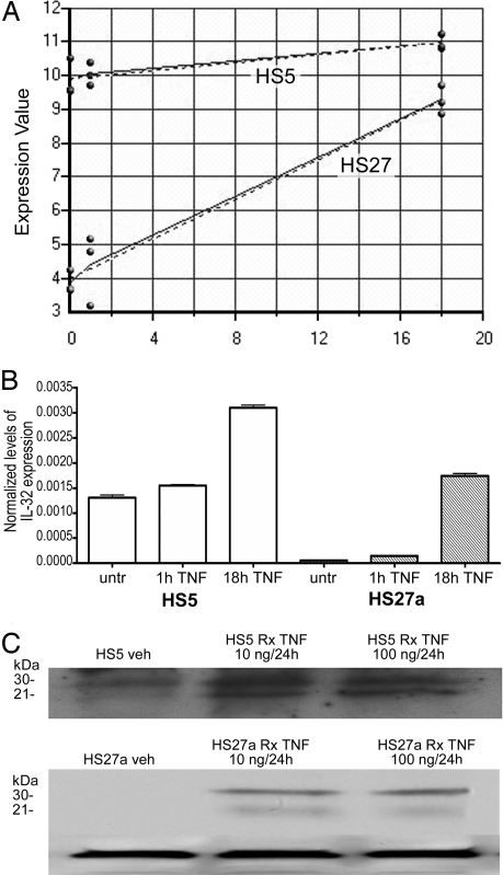
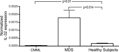
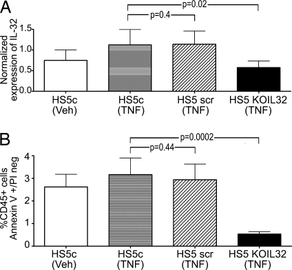
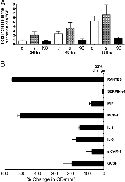
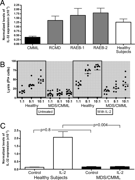
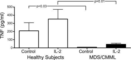
TNFalpha levels are elevated in the marrows of patients with myelodysplastic syndrome (MDS) and are associated with high rates of apoptosis, which contributes to hematopoietic failure. We observed that exposure of human marrow stroma cell lines HS5 and HS27a to TNFalpha increases levels of IL-32 mRNA. IL-32, in turn, induces TNFalpha. Marrow stroma from patients with MDS expressed 14- to 17-fold higher levels of IL-32 mRNA than healthy controls. In contrast, cells from patients with chronic myelomonocytic leukemia (CMML) expressed only one tenth the level of IL-32 measured in healthy controls. Human KG1a leukemia cells underwent apoptosis when cocultured with HS5 stromal cells, but knockdown of IL-32 in the stromal cells by using siRNA abrogated apoptosis in the leukemia cells. IL-32 knockdown cells also showed dysregulation of VEGF and other cytokines. Furthermore, CD56(+) natural killer cells from patients with MDS and CMML expressed IL-32 at lower levels than controls and exhibited reduced cytotoxic activity, which was unaffected by IL-2 treatment. We propose that IL-32 is a marrow stromal marker that distinguishes patients with MDS and CMML. Furthermore, IL-32 appears to contribute to the pathophysiology of MDS and may be a therapeutic target.


Conflict of interest statement

The authors declare no conflict of interest.

Figures

Fig. 1.
Fig. 1.
Expression of IL-32 in HS5 and HS27a cells in response to TNFα. (A) Log2 expression (y axis) of IL32 before (0) and 1 and 18 h after TNFα exposure (50 ng/ml) in HS5 and HS27a stroma cell lines (dotted lines, predicted expression; solid lines, measured expression). (B) IL-32 expression by quantitative RT-PCR before (untr) and 1 and 18 h after TNFα exposure (shown are results at 20 ng/ml TNF). (C) Western Blot of IL-32 protein in HS5 (Upper) and HS27a (Lower) cells. (Lower) β-actin served as control for both cell lines. Shown are IL-32 levels before treatment (Veh) and after treatment (Rx), with 10 ng/ml and 100 ng/ml of TNFα, respectively, for 24 h. The lower weight band likely represents IL-32α, and the higher likely represents IL-32γ.
Fig. 2.
Fig. 2.
Expression of IL-32 in primary marrow-derived stroma cells from untreated patients with CMML (n = 6) or MDS (n = 13) and healthy control subjects (n = 6). mRNA levels were determined by quantitative RT-PCR. P values are in comparison with healthy subjects. IL-32 levels were significantly lower in patients with CMML (P = 0.01) and higher in patients with MDS (P = 0.014).
Fig. 3.
Fig. 3.
IL-32 expression in HS5 stroma and effect on apoptosis in cocultured KG1a cells. (A) Expression of IL-32 in unmodified (HS5c) and modified (HS5 scr or HS5 KOIL32) stroma cells under control conditions (Veh) or after exposure to 100 ng/ml TNFα. IL-32 mRNA levels were determined by quantitative RT-PCR (n = 7; mean ± 1 SD). (B) Early stage apoptosis in KG1a cells in control cultures containing unmodified HS5 cells (HS5c) and in cocultures with HS5 cells transfected with either a scrambled siRNS sequence (HS5scr) or siRNA specific for IL-32 (HS5KOIL32). Cells were cultured in normal medium (Veh) or in the presence of 100 ng/ml TNFα. Apoptosis was determined by flow cytometry. Only CD45+ (KG1a) cells were considered.
Fig. 4.
Fig. 4.
Secretion of cytokines from HS5 stroma cells. Unmodified HS5 cells (c) or HS5 cells transfected with scrambled siRNA (s) or with IL-32-specific siRNA (KO) were used. (A) VEGF levels at 24, 48, and 72 h in the stroma cell supernatants by using ELISA. Changes in cytokine concentration are expressed as fold increase over control supernatants mean ± SD. (B) Expression of 36 cytokines in HS5KOIL32 cells in comparison with unmodified HS5 cells was determined by proteome profiler cytokine array (R&D Systems). Shown are changes (mean ± SD) in eight cytokines, which decreased significantly in HS5KOIL32 cells. The vertical line indicates a decrease of 33%. Levels were determined after 72 h of culture. The membrane contained probes for C5a, ICAM-1, IL-4, IL-13, IL-32α, MIP-1β, CD40 ligand, IFN-γ, IL-5, IL-16, IP-10, RANTES, G-CSF, IL-1α, IL-6, IL-17, I-TAC, SDF-1, GM-CSF, IL-1β, IL-8, IL-17E, MCP-1, Serpin-E1, GROα, IL-1ra, IL-10, IL-23, MIF, TNFα, I-309, IL-2, IL-12p70, IL-27 MIP-1α, and TREM-1.
Fig. 5.
Fig. 5.
IL-32 expression and NK activity. (A) IL-32 expression in peripheral blood CD56+ cells (n = 60). Shown are steady-state relative levels of expression of mRNA determined by quantitative RT-PCR. CMML, chronic myelomonocytic leukemia. MDS patients are shown by disease category [RCMD, refractory cytopenia with multilineage dysplasia; RAEB-1, refractory anemia with excess blasts type 1 (5–9% blasts); RAEB-2, refractory anemia with excess blasts, type 2 (10–19% blasts)]. Baseline expression of IL-32 in CD56+ cells from peripheral blood of patients with CMML was significantly lower than in CD56+ cells from healthy subjects (P = 0.001). Conversely, in patients with MDS, baseline expression of IL-32 tended to be higher than in healthy controls, although the difference did not reach statistical significance (P = 0.48). (B) Cytolytic function of NK cells. CD56+ cells were obtained from the peripheral blood of healthy subjects (n = 7) and patients with CMML or MDS (n = 13) and assayed at three different effector/target ratios (1:1; 5:1, and 10:1) against K562 cells. CD56+ cells were untreated or cultured for 24 h with 500 units/ml IL-2. Lytic activity was measured after 4 h of incubation of the effector cells (CD56+ cells) with target cells (K562). Lytic units were calculated from triplicate. Black dots show individual results, horizontal bars the mean percentage of lysis. (C) Expression of IL-32 in incubated peripheral blood CD56+ cells from healthy subjects (n = 7) and patients with MDS or CMML (because no difference was noted between MDS and CMML cells, results are lumped; n = 26). IL-32 levels were determined in unmodified cells (control) and cells that had been cultured with 500 units/ml IL-2 for 18 h. P values are for MDS/CMML patients in comparison with healthy subjects.
Fig. 6.
Fig. 6.
Secretion of TNFα by CD56+ NK cells from healthy subjects (n = 8) and patients with MDS or CMML (MDS; n = 16). TNFα was measured by ELISA at baseline (control) and 24 h after 500 units/ml IL-2. Shown is the mean ± SD (pg/ml). Although there was considerable variation in cells from healthy donors, little or no TNFα was secreted from MDS-derived cells even after activation by IL-2.

References

    1. Deeg HJ. In: Thomas' Hematopoietic Cell Transplantation. 4th Ed. Appelbaum FR, Forman SJ, Negrin RS, Blume KG, editors. Oxford: Blackwell; 2007.
    1. de Lima M, Giralt S. Allogeneic transplantation for the elderly patient with acute myelogenous leukemia or myelodysplastic syndrome. Semin Hematol. 2006;43:107–117. - PubMed
    1. Mundle SD, et al. Indication of an involvement of interleukin-1 beta converting enzyme-like protease in intramedullary apoptotic cell death in the bone marrow of patients with myelodysplastic syndromes. Blood. 1996;88:2640–2647. - PubMed
    1. Gersuk GM, et al. A role for tumor necrosis factor-α, Fas and Fas-Ligand in marrow failure associated with myelodysplastic syndrome. Br J Haematol. 1998;103:176–188. - PubMed
    1. Zang DY, Goodwin RG, Loken MR, Bryant E, Deeg HJ. Expression of tumor necrosis factor-related apoptosis-inducing ligand, Apo2L, and its receptors in myelodysplastic syndrome: effects on in vitro hemopoiesis. Blood. 2001;98:3058–3065. - PubMed

Publication types

MeSH terms