Comparative Study
. 2008 Feb 26;105(8):3134-9.
doi: 10.1073/pnas.0712338105. Epub 2008 Feb 19.

Acute regulation of aquaporin-2 phosphorylation at Ser-264 by vasopressin

Affiliations
Comparative Study

Acute regulation of aquaporin-2 phosphorylation at Ser-264 by vasopressin

Robert A Fenton et al. Proc Natl Acad Sci U S A. .

Abstract

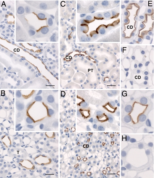
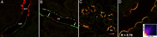
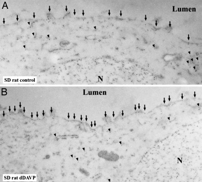
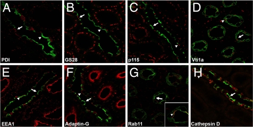
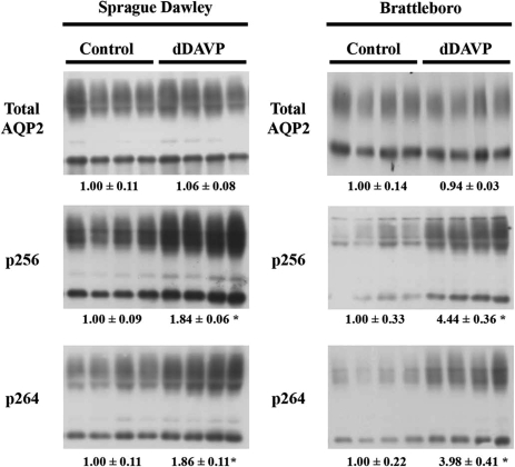
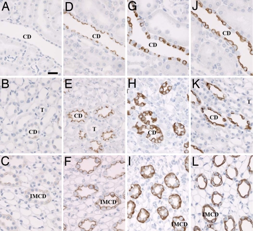
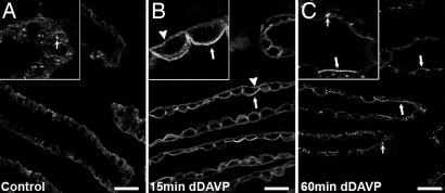
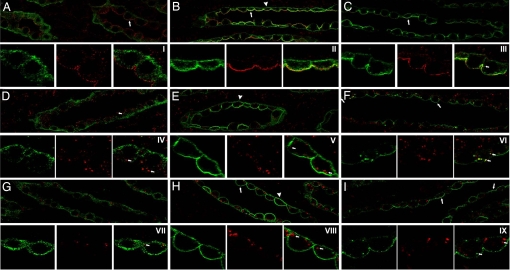
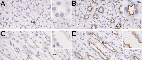
By phosphoproteome analysis, we identified a phosphorylation site, serine 264 (pS264), in the COOH terminus of the vasopressin-regulated water channel, aquaporin-2 (AQP2). In this study, we examined the regulation of AQP2 phosphorylated at serine 264 (pS264-AQP2) by vasopressin, using a phospho-specific antibody (anti-pS264). Immunohistochemical analysis showed pS264-AQP2 labeling of inner medullary collecting duct (IMCD) from control mice, whereas AQP2 knockout mice showed a complete absence of labeling. In rat and mouse, pS264-AQP2 was present throughout the collecting duct system, from the connecting tubule to the terminal IMCD. Immunogold electron microscopy, combined with double-labeling confocal immunofluorescence microscopy with organelle-specific markers, determined that the majority of pS264 resides in compartments associated with the plasma membrane and early endocytic pathways. In Brattleboro rats treated with [deamino-Cys-1, d-Arg-8]vasopressin (dDAVP), the abundance of pS264-AQP2 increased 4-fold over controls. Additionally, dDAVP treatment resulted in a time-dependent change in the distribution of pS264 from predominantly intracellular vesicles, to both the basolateral and apical plasma membranes. Sixty minutes after dDAVP exposure, a proportion of pS264-AQP2 was observed in clathrin-coated vesicles, early endosomal compartments, and recycling compartments, but not lysosomes. Overall, our results are consistent with a dynamic effect of AVP on the phosphorylation and subcellular distribution of AQP2.


Conflict of interest statement

The authors declare no conflict of interest.

Figures

Fig. 1.
Fig. 1.
Immunoperoxidase labeling of pS264–AQP2 in normal kidney. (A and B) Weak labeling of pS264 is evident in all regions of the CD in normal rat kidney [cortex (A) and inner stripe of outer medulla (B)]. (A and B Insets) At high magnification, labeling is predominantly apical membrane-associated, with some intracellular staining. (C and D) A similar pattern of labeling is observed in normal mouse kidney [cortex (C) and inner stripe of outer medulla (D)], with labeling restricted to the apical membrane domains (Insets). (E and F) AQP2 knockout mouse (F) shows a complete absence of p264 labeling compared with control mouse (E). (G and H) Preabsorption of anti-pS264 with nonphosphorylated peptide (G) or pS264 peptide (H) indicates that labeling is specific for the pS264–AQP2. T, thick ascending limb; PT, proximal tubule. (Scale bars, 20 μm.)
Fig. 2.
Fig. 2.
Confocal laser-scanning microscopy of pS264–AQP2 in normal rat kidney. (A) Double immunofluorescence labeling of pS264 (green) and calbindin (red) identified pS264 in connecting tubules. (B) Double immunofluorescence labeling of p264 (green, arrows) and [H+]ATPase (red) determined that pS264 is expressed in collecting duct principal cells. (C) Total AQP2 (red) and pS264 (green) colocalize (yellow) in collecting duct. (D) Higher magnification shows a high degree of correlation (depicted in histogram, inset) between total AQP2 and pS264 [Pearson correlation coefficient (R) in the colocalized volume, 0.78]. DCT, distal convoluted tubule.
Fig. 3.
Fig. 3.
Immunoelectron microscopy of pS264–AQP2 in normal rat kidney. (A) In rat kidney collecting duct principal cells, immunogold labeling of pS264 is observed intracellularly (arrowheads) and in the apical plasma membrane domains (arrows). (B) After dDAVP treatment (30 min), the number of gold particles increases in the apical plasma membrane (arrows), but intracellular gold can still be observed (arrowheads). N, nucleus.
Fig. 4.
Fig. 4.
Subcellular distribution of pS264–AQP2 in normal rat kidney collecting duct. For all images, pS264 is depicted in green, the specific intracellular marker is depicted in red, and the overlaid images show colocalized pixels in yellow, with arrows indicating pS264 and arrowheads indicating the intracellular compartment. Little colocalization is observed with the ER marker PDI (A), the medial Golgi marker GS28 (B), the cis Golgi marker p115 (C), and the TGN marker Vti1a (D). Some colocalization is observed in some tubules with the early endosome marker EEA1 (E), the clathrin-coated vesicle marker adaptin-G (F), and the recycling endosome marker Rab11 (G). No colocalization is observed with the lysosomal marker cathepsin D (H). See SI Table 1 for statistical comparison.
Fig. 5.
Fig. 5.
The abundance of pS264–AQP2 is increased with short-term vasopressin exposure. Immunoblots assessing relative total AQP2, pS256, and pS264 abundance in inner medulla homogenates from Sprague–Dawley rats or Brattleboro rats after dDAVP treatment (30 min). Each lane was loaded with a sample from a different rat. The values are mean band densities normalized such that the mean for the control rats is defined as 1. *, Significant change in mean band densities among groups.
Fig. 6.
Fig. 6.
Short-term vasopressin exposure increases pS264–AQP2 abundance at the plasma membrane. (A–C) Weak intracellular labeling of pS264 is evident in Brattleboro rat kidney cortex (A), inner stripe of outer medulla (B), and inner medulla (C). (D–F) After 30 min of dDAVP exposure, pS264 labeling increases in intensity predominantly at the apical plasma membrane, although weak basolateral plasma membrane labeling is apparent in the IMCD (F). (G–L) In comparison, total AQP2 is highly abundant in all regions before dDAVP exposure (G–I) but translocates to the apical membrane upon stimulation (J–L). (Scale bar, 10 μm.)
Fig. 7.
Fig. 7.
Short-term vasopressin exposure results in increased pS264–AQP2 in the basolateral and apical membranes. (A) In control Brattleboro rats, weak intracellular labeling of pS264 is observed. In some tubules, at high magnification (Inset) labeling is localized to small vesicles (small arrows). (B) After 15 min of dDAVP treatment, pS264 labeling increases on the basolateral (arrowheads) and apical membrane (arrows); this is more apparent at high magnification (Inset). (C) After 60 min, pS264 is predominantly associated with the apical plasma membrane (arrows) and small distinct intracellular vesicles (small arrows). (Scale bars, 20 μm.)
Fig. 8.
Fig. 8.
Subcellular distribution of pS264–AQP2 after short-term vasopressin treatment. Brattleboro rats were treated with vehicle (15 or 60 min) or dDAVP (15 or 60 min), and double immunofluorescence labeling was performed. For all images, pS264 is depicted in green, the specific intracellular marker is depicted in red, and the overlaid images show colocalized pixels is in yellow. (A) Weak intracellular pS264 labeling is apparent in controls that does not colocalize with the clathrin-coated pit marker adaptin-β (arrow). (B) pS264 labeling increases on the basolateral (arrowheads) and apical membrane (arrows) after 15 min of dDAVP. (C) After 60 min, pS264 is predominantly associated with the apical plasma membrane (arrows) and small intracellular vesicles. At high magnification, this series of events is more apparent (I–III) with distinct pS264 labeling of vesicle structures at 60 min (small arrows). (D) pS264 labeling does not colocalize with the early endosome marker EEA1. (E) pS264 labeling increases on the basolateral (arrowheads) and apical membrane after 15 min of dDAVP but does not colocalize with EEA1. (F) After 60 min, pS264 is associated with the apical plasma membrane (arrows) and small EEA1-positive intracellular vesicles. At high magnification (IV–VI), pS264 labeling of both EEA1-positive vesicles (small arrows) and EEA1-negative vesicles is apparent after 60 min. (G–I) pS264 labeling does not colocalize at any time point with the lysosome marker cathepsin D. At high magnification (VII–IX), distinct pS264-positive or cathepsin D-positive vesicles are apparent (small arrows).
Fig. 9.
Fig. 9.
Long-term increases in circulating vasopressin levels result in predominantly apical plasma membrane localization of pS264–AQP2. (A and B) Weak intracellular labeling of pS264 is evident in control Brattleboro rat kidney inner medulla (A) that increases in abundance at the apical plasma membrane after 5 days of continuous dDAVP infusion (B). (C and D) Weak intracellular pS264 labeling is apparent in the inner medulla of water-loaded Sprague–Dawley rats (C) that increases in abundance at the apical plasma membrane after 5 days of water restriction (D).

References

    1. Nielsen S, et al. Aquaporins in the kidney: from molecules to medicine. Physiol Rev. 2002;82:205–244. - PubMed
    1. Fushimi K, Sasaki S, Marumo F. Phosphorylation of serine 256 is required for cAMP-dependent regulatory exocytosis of the aquaporin-2 water channel. J Biol Chem. 1997;272:14800–14804. - PubMed
    1. Katsura T, Gustafson CE, Ausiello DA, Brown D. Protein kinase A phosphorylation is involved in regulated exocytosis of aquaporin-2 in transfected LLC-PK1 cells. Am J Physiol. 1997;272:F817–F822. - PubMed
    1. Kamsteeg EJ, Heijnen I, van Os CH, Deen PM. The subcellular localization of an aquaporin-2 tetramer depends on the stoichiometry of phosphorylated and nonphosphorylated monomers. J Cell Biol. 2000;151:919–930. - PMC - PubMed
    1. McDill BW, et al. Congenital progressive hydronephrosis (cph) is caused by an S256L mutation in aquaporin-2 that affects its phosphorylation and apical membrane accumulation. Proc Natl Acad Sci USA. 2006;103:6952–6957. - PMC - PubMed

Publication types