Comparative Study
. 2008 Feb 26;105(8):2800-5.
doi: 10.1073/pnas.0711963105. Epub 2008 Feb 19.

Quantitative biochemical rationale for differences in transmissibility of 1918 pandemic influenza A viruses

Affiliations
Comparative Study

Quantitative biochemical rationale for differences in transmissibility of 1918 pandemic influenza A viruses

Aravind Srinivasan et al. Proc Natl Acad Sci U S A. .

Abstract

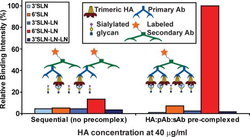
The human adaptation of influenza A viruses is critically governed by the binding specificity of the viral surface hemagglutinin (HA) to long (chain length) alpha2-6 sialylated glycan (alpha2-6) receptors on the human upper respiratory tissues. A recent study demonstrated that whereas the 1918 H1N1 pandemic virus, A/South Carolina/1/1918 (SC18), with alpha2-6 binding preference transmitted efficiently, a single amino acid mutation on HA resulted in a mixed alpha2-3 sialylated glycan (alpha2-3)/alpha2-6 binding virus (NY18) that transmitted inefficiently. To define the biochemical basis for the observed differences in virus transmission, in this study, we have developed an approach to quantify the multivalent HA-glycan interactions. Analysis of the molecular HA-glycan contacts showed subtle changes resulting from the single amino acid variations between SC18 and NY18. The effect of these changes on glycan binding is amplified by multivalency, resulting in quantitative differences in their long alpha2-6 glycan binding affinities. Furthermore, these differences are also reflected in the markedly distinct binding pattern of SC18 and NY18 HA to the physiological glycans present in human upper respiratory tissues. Thus, the dramatic lower binding affinity of NY18 to long alpha2-6 glycans, as against a mixed alpha2-3/6 binding, correlates with its inefficient transmission. In summary, this study establishes a quantitative biochemical correlate for influenza A virus transmission.


Conflict of interest statement

The authors declare no conflict of interest.

Figures

Fig. 1.
Fig. 1.
Molecular interactions of SC18, NY18, and AV18 HAs with α2-3 and α2-6. The glycan binding site on HA is shown with the critical amino acids including the highly conserved Neu5Ac anchors (Thr-136, Trp-153, Thr-155, and Leu-194). Tyr-95 and His-183 are not shown for clarity. The amino acid positions are numbered based on H1N1 HA. (A) Shown is the interaction of the SC18 HA with long α2-6 in the umbrella-like topology. Lys-222, Asp-225, and Gln-226 provide critical contacts with the base region, and Asp-190, Gln-192, and Ser-193 provide optimal contacts with the extension region. (B) Interaction of NY18 HA with the long α2-6 shows the loss of the critical contact of Asp-225 with the base region (as observed in the case of SC18 HA in A). (C) Interaction of AV18 HA with α2-3 in cone-like topology shows that HA interactions with cone-like topology involves contacts only with the Neu5Ac and Gal sugars at the base in contrast to that of the umbrella-like topology of long α2-6. Glu-190 and Gln-226 in AV18 are positioned to provide optimal contacts with these sugars. The side-chain conformation of Glu-190 in AV18 was assigned based on that of Glu-190 in APR34 HA–α2-3 cocrystal structure. (D) The differences in the key amino acid positions between SC18, NY18, and AV18 HAs in comparison with the ASI30 and APR34 HAs. The differences in the amino acid contacts with α2-3 and α2-6 are quantified by using buried solvent-accessible area calculations [supporting information (SI) Fig. 6].
Fig. 2.
Fig. 2.
Binding assay to capture multivalent HA–glycan interactions. Shown is a comparison of binding signals between sequential binding assay and precomplexation of HA units with primary (pAb) and labeled secondary (sAb) antibodies. The conditions of the sequential assay (see Materials and Methods) favor the formation of HA/pAb/sAb in a 1:1:1 ratio. Despite the abundance of the labeled sAb (1 per HA unit) the minimal binding signals observed even at a high HA concentration of 40 μg/ml supports the low affinity binding of a single glycan to a HA unit (cyan circle). On the other hand, the conditions for precomplexation of HA/pAb/sAb in a ratio of 4:2:1 favor the spatial arrangement of four HA units per precomplex. This spatial arrangement enhances the glycan binding signals via multivalency as shown by at least an 8-fold increase in binding to 6′SLN-LN given that there are four binding events (each event shown by a cyan circle) per precomplexed HA unit.
Fig. 3.
Fig. 3.
Dose-dependent direct glycan binding of SC18, NY18, and AV18 HA. (A–C) The binding signals (expressed as percentage of maximum) of SC18, NY18, and AV18, respectively, in the dose-dependent direct binding assay. SC18 HA shows binding signals only to the long α2-6. At saturating HA concentrations (40 μg/ml), both SC18 and NY18 HA show identical binding to long α2-6, and NY18 HA also shows high binding signals to long α2-3. On the other hand, AV18 HA shows a reverse trend (in comparison with SC18 HA) of high-affinity α2-3 binding and minimal α2-6 binding. (D) Comparison of the long α2-6 (6′SLN-LN) binding of SC18, TX91, and Mos99 HAs with that of NY18 HA. Whereas the long α2-6 binding signal of NY18 HA approaches that of the other HAs at high concentrations, the binding curve of NY18 HA over the entire concentration range indicates that it has a dramatically lower long α2-6 binding affinity. Human-adapted SC18 and TX91 H1N1 viruses transmit efficiently in the ferret model (12). Human-adapted Mos99 H3N2 virus is a vaccine strain.
Fig. 4.
Fig. 4.
Binding of SC18 and NY18 HA to human tracheal sections. The apical side of the tracheal epithelia is indicated by white arrows. The binding pattern of SC18 HA is localized around specific regions on the apical side; on the other hand, NY18 HA shows well distributed apical binding pattern (HA in green against PI in red). Furthermore, NY18 HA also shows significant staining on the inner regions of the epithelia that express α2-3 glycans (indicated by MAL-II-staining patterns) (data not shown). (Magnification: ×25.)
Fig. 5.
Fig. 5.
Human tracheal costaining of SC18 and NY18 HA with Jacalin. The apical side of the tracheal epithelia is indicated by white arrows. The distinct apical binding patterns of SC18 and NY18 (in Fig. 4) are further elaborated by costaining the HAs (red) with Jacalin (green), a marker for goblet cells. The images were taken at ×25 magnification, and representative regions of the apical side are enlarged for clarity. The significant costain of SC18 HA with Jacalin (yellow) indicates that SC18 HA predominantly binds to goblet cells on the apical side of the epithelia. On the other hand, the binding pattern of NY18 HA has minimal overlap with that of Jacalin, indicating that it does not bind to goblet cells on the apical side of tracheal tissue.

References

    1. Tumpey TM, et al. Characterization of the reconstructed 1918 Spanish influenza pandemic virus. Science. 2005;310:77–80. - PubMed
    1. Kuiken T, et al. Host species barriers to influenza virus infections. Science. 2006;312:394–397. - PubMed
    1. Russell RJ, et al. Avian and human receptor binding by hemagglutinins of influenza A viruses. Glycoconj J. 2006;23:85–92. - PubMed
    1. Shinya K, et al. Avian flu: Influenza virus receptors in the human airway. Nature. 2006;440:435–436. - PubMed
    1. Skehel JJ, Wiley DC. Receptor binding and membrane fusion in virus entry: The influenza hemagglutinin. Annu Rev Biochem. 2000;69:531–569. - PubMed

Publication types

Substances