Comparative Study
. 2008 Feb 26;105(8):2883-8.
doi: 10.1073/pnas.0711983105. Epub 2008 Feb 15.

Generation of human induced pluripotent stem cells from dermal fibroblasts

Affiliations
Comparative Study

Generation of human induced pluripotent stem cells from dermal fibroblasts

W E Lowry et al. Proc Natl Acad Sci U S A. .

Abstract

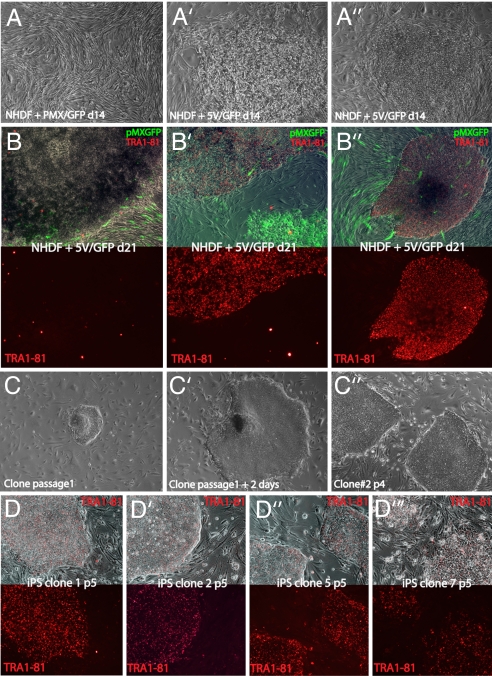
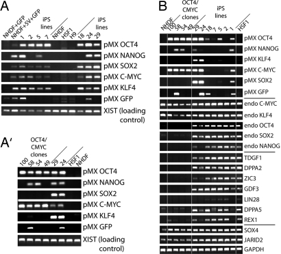
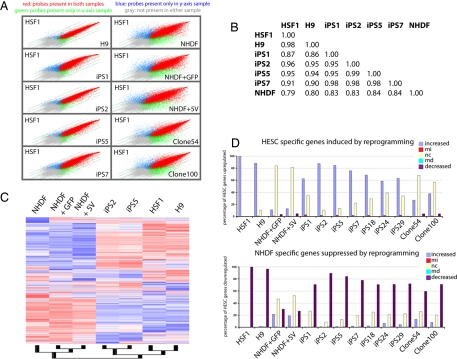
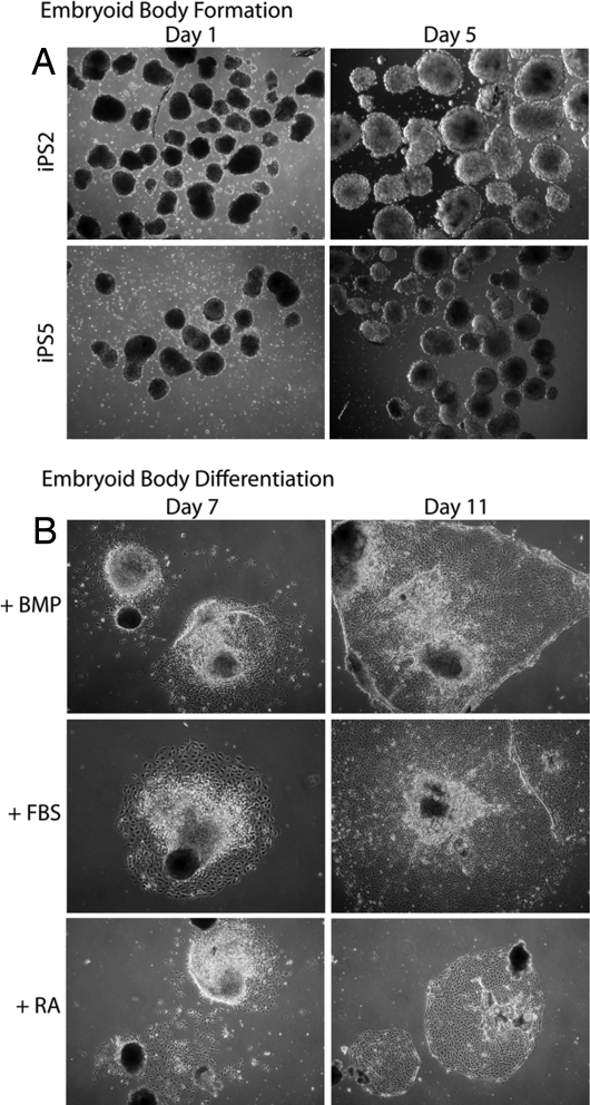
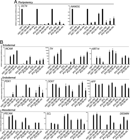
The generation of patient-specific pluripotent stem cells has the potential to accelerate the implementation of stem cells for clinical treatment of degenerative diseases. Technologies including somatic cell nuclear transfer and cell fusion might generate such cells but are hindered by issues that might prevent them from being used clinically. Here, we describe methods to use dermal fibroblasts easily obtained from an individual human to generate human induced pluripotent stem (iPS) cells by ectopic expression of the defined transcription factors KLF4, OCT4, SOX2, and C-MYC. The resultant cell lines are morphologically indistinguishable from human embryonic stem cells (HESC) generated from the inner cell mass of a human preimplantation embryo. Consistent with these observations, human iPS cells share a nearly identical gene-expression profile with two established HESC lines. Importantly, DNA fingerprinting indicates that the human iPS cells were derived from the donor material and are not a result of contamination. Karyotypic analyses demonstrate that reprogramming of human cells by defined factors does not induce, or require, chromosomal abnormalities. Finally, we provide evidence that human iPS cells can be induced to differentiate along lineages representative of the three embryonic germ layers indicating the pluripotency of these cells. Our findings are an important step toward manipulating somatic human cells to generate an unlimited supply of patient-specific pluripotent stem cells. In the future, the use of defined factors to change cell fate may be the key to routine nuclear reprogramming of human somatic cells.


Conflict of interest statement

The authors declare no conflict of interest.

Figures

Fig. 1.
Fig. 1.
iPS clones share HESC morphology. (A′) Heterogeneous morphology of colonies of NHDF1 infected with empty pMX virus and GFP-containing pMX viruses in a 5:1 ratio (pMX/GFP) or a combination of six pMX viruses each carrying one of the five defined transcription factors or GFP (5V/GFP), at day 14 after infection in phase contrast. (B–B″) Phase-contrast images of different colonies from 5V/GFP transduced cultures merged with live TRA-1–81 staining (red) and GFP fluorescence derived from the pMX-GFP virus (green) (Upper) and the TRA-1–81 channel separately (Lower) from cultures transduced with 5V/GFP. Note that only a minor proportion of colonies are TRA-1–81-positive as seen in B and B′. The staining in TRA-1–81-positive colonies was indistinguishable from that obtained with HESC (data not shown). (C–C″) Phase-contrast images of iPS clones at different passages. (D–D‴) “Live” Tra-1–81 staining and merge with phase-contrast appearance of indicated iPS clones at passage 5.
Fig. 2.
Fig. 2.
iPS clones express key HESC markers. (A and A′) PCR for retroviral integration events on genomic DNA derived from iPS and “early” OCT4/C-MYC clones, control NHDF1, NHDF1 cells infected with control (GFP) or defined factor viruses (5V + GFP), and HSF1 or H9 HESC, with primers that specifically recognize each of the integrated viruses. Loading control: PCR for a genomic region on the X chromosome within the XIST locus. iPS clones 24 and 29 are included in A′ as a positive control for the PCR conditions. (B) RT-PCR for pMX retroviral transcription and expression of endogenous counterparts of the defined factors as well as additional HESC-specific genes (TDGF1 through REX1) in iPS clones, NHDF1 and the HSF1 HESC, and in OCT4/CMYC clones. Note that iPS24 and 29 and the OCT4/CMYC clones, respectively, largely failed to suppress expression from the viruses they received.
Fig. 3.
Fig. 3.
The transcriptome of iPS clones is highly similar to that of HESC. (A) Scatter-plot presentation of the expression values for all probe sets derived from genome-wide microarray expression data of indicated cell types. NHDF1 + GFP and NHDF1 + 5V denote a pool of fibroblasts infected with pMX/pMX-GFP control viruses or viruses carrying the five defined factors plus GFP at day 18 after infection. Like the H9 HESC line, iPS clones 2 and 5 appear highly similar to the HSF1 HESC, whereas iPS lines 1 and 7 appear slightly less similar to HESC. (B) Global Pearson correlation of the entire expression data (from Affymetrix microarrays) between indicated cell types. (C) Hierarchical clustering of gene-expression data of the indicated cell types. Normalization and expression analysis was performed with DNA-chip analyzer (dChip). A 20% presence call was used to filter genes for clustering, and redundant probe sets were removed. (D) The 2,000 most up- and down-regulated genes in HSF1 versus NHDF were determined from genome-wide expression datasets and analyzed for up-regulation, down-regulation, or no change in expression between iPS clones or pools of infected NHDF cells and NHDF. MI and MD denote statistically marginal increase or decrease, respectively.
Fig. 4.
Fig. 4.
iPS cells form embryoid bodies similarly to HESCs. (A) Phase-contrast images of EBs generated from iPS clones 2 and 5. (B) Growth of iPS-derived EBs upon plating onto adherent tissue culture dishes under three different media conditions. BMP, bone morphogenetic protein 4.
Fig. 5.
Fig. 5.
Pluripotency of iPS cells and up-regulation of ectodermal, endodermal, and mesodermal markers. (A) Real-time RT-PCR analysis of pluripotency gene expression in iPS and control HESC (HSF1) induced to differentiate by EB formation and subsequent plating under indicated conditions [BMP4, FBS, retinoic acid (RA)] relative to GAPDH expression. The y axis represents relative fold change upon differentiation. Note that EB differentiation induces down-regulation of pluripotency markers such as OCT4 and NANOG. (B) As in A except that expression of marker genes for different germ layers was analyzed. The specificity of each marker for a given germ layer is indicated. The y axis represents relative fold of induction over undifferentiated cells. Note that although the degree of induction of lineage markers is sometimes variable between HESC and iPS clones, the pattern is consistent.

References

    1. Byrne JA, et al. Producing primate embryonic stem cells by somatic cell nuclear transfer. Nature. 2007;450:497–502. - PubMed
    1. Takahashi K, Yamanaka S. Induction of pluripotent stem cells from mouse embryonic and adult fibroblast cultures by defined factors. Cell. 2006;126:663–676. - PubMed
    1. Okita K, Ichisaka T, Yamanaka S. Generation of germline-competent induced pluripotent stem cells. Nature. 2007;448:313–317. - PubMed
    1. Wernig M, et al. In vitro reprogramming of fibroblasts into a pluripotent ES cell-like state. Nature. 2007;448:318–324. - PubMed
    1. Maherali N, et al. Directly reprogrammed fibroblasts show global epigenetic remodeling and widespread tissue contribution. Cell Stem Cell. 2007;1:55–70. - PubMed

Publication types

MeSH terms

Associated data