. 2008 Apr 18;283(16):10835-47.
doi: 10.1074/jbc.M710363200. Epub 2008 Feb 20.

Transforming growth factor-beta1 (TGFbeta1) stimulates connective tissue growth factor (CCN2/CTGF) expression in human gingival fibroblasts through a RhoA-independent, Rac1/Cdc42-dependent mechanism: statins with forskolin block TGFbeta1-induced CCN2/CTGF expression

Affiliations

Transforming growth factor-beta1 (TGFbeta1) stimulates connective tissue growth factor (CCN2/CTGF) expression in human gingival fibroblasts through a RhoA-independent, Rac1/Cdc42-dependent mechanism: statins with forskolin block TGFbeta1-induced CCN2/CTGF expression

Samuel A Black Jr et al. J Biol Chem. .

Abstract

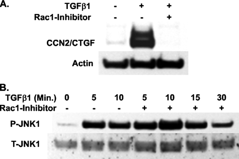
Regulation of connective tissue growth factor (CCN2/CTGF) in gingival fibroblasts is unique and may provide therapeutic opportunities to treat oral fibrotic diseases. RhoA was previously implicated in mediating the expression of CCN2/CTGF. We now present evidence that Rho family GTPases Rac1 and Cdc42 are the principal mediators of the transforming growth factor-beta1 (TGFbeta1)-stimulated expression of CCN2/CTGF in primary human gingival fibroblasts. TGFbeta1 does not stimulate RhoA activation in gingival fibroblasts, and the overexpression of dominant-negative RhoA does not reduce CCN2/CTGF expression in response to TGFbeta1. In contrast, the overexpression of dominant-negative forms of Cdc42 or Rac1 results in a dramatic reduction of CCN2/CTGF protein levels. Lovastatin and a geranylgeranyltransferase inhibitor reduce the TGFbeta1-stimulated levels of CCN2/CTGF protein by approximately 75 and 100%, respectively. We previously demonstrated that JNK1 phosphorylation by TGFbeta1 is also critical for TGFbeta1-induced CCN2/CTGF expression, and forskolin partially reduces levels of phosphorylated JNK1. Inhibition of geranylgeranyltransferase has no effect on levels of JNK phosphorylation in response to TGFbeta1 suggesting Rho-GTPases act independently of JNK1. The combination of lovastatin and forskolin results in a greater inhibitory effect than each agent alone and reduces CCN2/CTGF mRNA and protein expression by greater than 90%. This novel combination has additive inhibitory effects on the TGFbeta1-stimulated expression of CCN2/CTGF in human gingival fibroblasts through the simultaneous disruption of Rho- and JNK1-mediated pathways, respectively. This combination of available therapeutic compounds may therefore be useful in designing treatment strategies for oral fibrotic conditions in which gingival CCN2/CTGF is elevated.


Figures

FIGURE 1.
FIGURE 1.
The kinetics of the TGFβ1- and LPA-stimulated expression of CCN2/CTGF mRNA and protein levels differ in primary human gingival fibroblasts. Near confluent cultures of primary gingival fibroblasts were serum-starved for 12 h and then treated with 10 μm LPA, 5 ng/ml TGFβ1, or both for 2 or 6 h prior to harvest of total RNA or protein. A, real time qPCR analysis of the fold change in CCN2/CTGF mRNA expression versus no treatment control cultures. B, Western blot analysis of CCN2/CTGF protein expression. Each experiment was conducted at least twice with identical results. Data are represented as the mean ± S.D. and statistical evaluation per each experimental condition determined versus control, no treatment, cultures. *, p < 0.001; **, p < 0.05; #, p < 0.01.
FIGURE 2.
FIGURE 2.
TGFβ1 and LPA stimulate comparable levels of phospho-JNK1 in human gingival fibroblasts. Gingival fibroblast cultures serum-starved for 12 h were treated with 10 μm LPA, 5 ng/ml TGFβ1, or both for times indicated prior to harvest of total cell layer protein. A, representative Western blot of phosphorylated JNK1 in response to LPA or TGFβ1. B, densitometric analysis from three separate experiments showing no significant difference between the TGFβ1- and LPA-stimulated inductions of phosphorylated JNK1. Data are represented as the mean ± S.D. and statistical evaluation per each experimental condition determined versus control, no treatment, cultures. C, Western blot of JNK1 protein phosphorylation in response to TGFβ1 alone or in combination with LPA.
FIGURE 3.
FIGURE 3.
LPA is significantly less effective than TGFβ1 in stimulating the nuclear localization of phosphorylated Smad3. Gingival fibroblasts were serum-starved for 12 h and then treated with 5 ng/ml TGFβ1 and/or 10 μm LPA for times indicated. Nuclei were harvested and purified, and total nuclear protein was retained for Western blot analysis of nuclear phospho-Smad3. A, Western blot for α-tubulin (cytoplasmic marker) and lamin B (nuclear marker) demonstrating that purified nuclear extracts used in these experiments were free of cytoplasmic proteins. B, Western blot demonstrating nuclear levels of phospho-Smad3 following treatment with TGFβ1 for the times indicated. C, Western blot of nuclear phospho-Smad3 in response to TGFβ1 and LPA or both. Each experiment was conducted at least twice with the same outcomes. B and C, lamin B, a nuclear protein, was used as the loading control.
FIGURE 4.
FIGURE 4.
Lovastatin and forskolin work in concert to completely block the TGFβ1-induced expression of CCN2/CTGF in human gingival fibroblasts. A, Western blot of CCN2/CTGF protein levels in response to treatment with 20 μm of either lovastatin or simvastatin. Cell cultures were pretreated with lovastatin or simvastatin for 12 h. Media were then aspirated and replaced with media containing lovastatin or simvastatin combined with 5 ng/ml TGFβ1 and incubated for 6 h prior to harvest of total cell layer protein for Western blot analysis of CCN2/CTGF. B, Western blot of CCN2/CTGF protein in response to 20 μm lovastatin and 20 μm mevalonate. Cultures were pretreated with lovastatin for 12 h. Media were then aspirated and replaced with media containing lovastatin plus 20 μm mevalonate and 5 ng/ml TGFβ1. Cultures were incubated for an additional 6 h prior to harvest of total cell layer protein for Western blot analysis of CCN2/CTGF. C, Western blot of the TGFβ1-stimulated expression of CCN2/CTGF and the effects of 20 μm GGTI or FTI inhibitors. Cultures were pretreated with 20 μm GGTI or FTI for 12 h. Media were then aspirated, and GGTI or FTI was combined with 5 ng/ml TGFβ1. Cultures were then incubated for 6 h prior to harvest of total cell layer protein for Western blot analysis of CCN2/CTGF. D, Western blot of phosphorylated JNK1 levels in response to 5 ng/ml TGFβ1 and 20 μm GGTI. Cell cultures were pretreated with GGTI-supplemented media for 12 h. Media were then aspirated, and 20 μm GGTI was combined with 5 ng/ml TGFβ1, and cells were harvested at intervals, as indicated. E, real time qPCR analysis of CCN2/CTGF mRNA expression in response to challenge with 10 μm forskolin, 20 μm lovastatin, or both. Cell cultures were pretreated with 20 μm lovastatin (12 h) or 10 μm forskolin (1 h). Media were then aspirated and either lovastatin or forskolin was combined with 5 ng/ml TGFβ1 and incubated for an additional 4 h prior to harvest of total mRNA for real time qPCR of CCN2/CTGF mRNA. In experiments combining lovastatin and forskolin, cultures were pretreated with 20 μm lovastatin for 11 h. Medium was aspirated and replaced with medium containing 20 μm lovastatin and 10 μm forskolin and incubated for 1 h. Culture medium was again replaced with lovastatin and forskolin containing 5 ng/ml TGFβ1. Cultures were incubated for an additional 4 h prior to isolation of total RNA for real time qPCR analysis; *, p < 0.0001; **, p < 0.000005 compared with TGF-β1-stimulated cells. F, Western blot analysis demonstrating the strong combined inhibitory effect of lovastatin and forskolin on the TGFβ1-induced expression of CCN2/CTGF protein. Cultures were treated as described in E, but the final incubation time was 6 h prior to the harvest of total cell layer protein for Western blot analysis of CCN2/CTGF. Each experiment was repeated at least twice with the same outcomes. Representative blots are shown. Real time qPCR experiments were conducted twice in triplicate, and data are represented as the mean ± S.D. and statistical evaluation per each experimental condition determined versus control, no treatment, cultures.
FIGURE 5.
FIGURE 5.
Lovastatin is equally potent at inhibiting the TGFβ1-stimulated expression of CCN2/CTGF at concentrations of 10 and 20 μm. Cultures of primary human gingival fibroblasts were grown as described above. Cells were then treated for 12 h with increasing concentrations of lovastatin for 12 h or for 12 or 24 h with either 10 or 20 μm lovastatin. Culture media were then aspirated and replaced with equivalent concentrations of lovastatin and 5 ng/ml TGFβ1. Cells were incubated for an additional 6 h prior to harvest of total cell layer protein. The effects of the duration of the inhibition with lovastatin and the concentration on the TGFβ1-induced (5 ng/ml) expression of CCN2/CTGF protein were assessed using Western blot analysis. A, Western blot showing the effects of different concentrations of lovastatin on the TGFβ1-induced expression of CCN2/CTGF protein. B, representative Western blot of the effects of different concentrations of lovastatin and incubation times on CCN2/CTGF protein. C, densitometric analysis of CCN2/CTGF protein levels normalized to total β-actin. Statistical significance was determined comparing lovastatin-treated lanes to those treated with TGFβ1 alone (*, p < 0.0001). Experiments were repeated at least twice with the same findings.
FIGURE 6.
FIGURE 6.
The TGFβ1-stimulated expression of CCN2/CTGF is inhibited in gingival cells transfected with the C3 exotransferase of C. botulinum. Gingival fibroblast cultures were grown to near confluence and then placed in serum-free media containing the Chariot small protein transfection reagent and 5 μg of C3 protein. Cultures were incubated for 2 h, and media were replaced with fresh, serum-free media and incubated for 10 h. 5 ng/ml TGFβ1 was then added to cultures with or without C3 and incubated for an additional 6 h prior to harvest of total cell protein. A, representative Western blot demonstrating the inhibition of C3 on the TGFβ1-induced expression of CCN2/CTGF. The ROCK-specific inhibitor Y27632 blocks elevations in CCN2/CTGF protein levels stimulated by TGFβ1 without reducing levels of phosphorylated JNK1. B, Western blot of CCN2/CTGF protein showing the inhibitors Y27632 and H89 block the TGFβ1-induced expression of CCN2/CTGF in gingival cells. Serum-starved cultures of gingival fibroblasts were pretreated with 10 μm of either Y27632 or H89 for 1 h prior to the addition of 5 ng/ml TGFβ1. After a subsequent 6-h incubation, total protein was collected for Western blot analyses of CCN2/CTGF protein. Western blot of phosphorylated JNK1 showing that the inhibition of CCN2/CTGF by Y27632 (C) or H89 (D) was not because of the reduction of JNK1 activation. Cultures were pretreated with either 10 μm Y27632 or H89 for 1 h. Medium was then replaced with fresh medium containing an equivalent concentration of inhibitor and 5 ng/ml TGFβ1 and incubated for times indicated prior to harvest of total protein for analysis of phosphorylated JNK. Each experiment was conducted at least twice with the same results. E, LPA, not TGFβ1, stimulates the activation of RhoA in primary human gingival fibroblasts. Gingival fibroblasts were serum-starved and then treated with either 5 ng/ml TGFβ1, 10 μm LPA, 1 μm PGE2, or 10 μm forskolin for 3 min. One set of cultures was treated with the combination of TGFβ1 and LPA. Cells were collected for pulldown assay for activated RhoA. GDP and GTPγS were used as negative and positive controls, respectively.
FIGURE 7.
FIGURE 7.
Inhibition of Rac1 activation leads to the complete loss of TGFβ1-induced CCN2/CTGF expression without interfering with JNK1 phosphorylation. Gingival fibroblasts were serum-starved and then pretreated with 100 μm Rac1 inhibitor for 1 h. Cultures were then treated with 5 ng/ml TGFβ1 for 6 h prior to harvest of total cellular protein Western blot of CCN2/CTGF protein expression (A) or for times indicated for assessment of phosphorylation of JNK1 by Western blot (B). The experiment was performed twice with the same results.
FIGURE 8.
FIGURE 8.
Overexpression of dominant-negative Rac1 or Cdc42, and not RhoA, leads to the near complete loss of CCN2/CTGF expression induced by TGFβ1 in gingival cells. Gingival cell cultures were infected with recombinant adenovirus (Ad) expressing dominant-negative forms of either Rac1, RhoA, Ras, or Cdc42 and incubated for 48 h. Cells were then fed with media containing 5 ng/ml TGFβ1. Cultures were incubated for an additional 6 h, and total cell layer protein was harvested for Western blot analysis of CCN2/CTGF protein levels. A, levels of recombinant adenovirus expressing β-galactosidase were used to optimize viral load in primary gingival fibroblasts. Near 100% infection of cells in culture was achieved with no obvious alteration in cell morphology. The same titer of each dominant-negative (DN) virus was used to infect cultures. B, representative Western blot of CCN2/CTGF protein levels in response to the overexpression of dominant-negative GTPases. C, densitometric analysis of Western blot data obtained from three separate experiments. Bars represent the mean CCN2/CTGF protein levels normalized to total β-actin, ±S.D., and statistical evaluation per each experimental condition determined versus control, Ad-β-galactosidase-infected (Ad-β-Gal) cultures (*, p < 0.001; **, p < 0.05; ***, p < 0.0005).
FIGURE 9.
FIGURE 9.
Several signaling pathways work together in the TGFβ1-induced expression of CCN2/CTGF in primary human gingival fibroblasts. Cdc42, Rac1, and Ras are small GTPases involved in the TGFβ1-stimulated expression of CCN2/CTGF whose activity relies upon the availability of products of the cholesterol biosynthesis pathway. Proposed stimulation of protease secretion in response to stimulation with TGFβ1 may result in the activation of protease-activated receptors and ultimately lead to the activation of Ras to prolong CCN2/CTGF expression in these cells (hypothetical pathway indicated by broken arrows). TGFβ1 and LPA both stimulate CCN2/CTGF expression in gingival cells with TGFβ1 being the more potent inducer. Both TGFβ1 and LPA stimulate the activating phosphorylation of JNK1 and nuclear localization of phosphorylated Smad3. JNK1 does not, however, enhance Smad3 nuclear localization in gingival fibroblasts but is required for TGFβ1 stimulation of CCN2/CTGF expression (40). LPA induces only a weak nuclear localization of Smad3. Data support that although LPA stimulates signaling pathways also stimulated by TGFβ1, LPA and TGFβ1 each uniquely stimulate a subset of complementary parallel signaling events that result in additive up-regulation of CCN2/CTGF levels in gingival fibroblasts.

References

    1. Bork, P. (1993) FEBS Lett. 327 125-130 - PubMed
    1. Oemar, B. S., and Luscher, T. F. (1997) Arterioscler. Thromb. Vasc. Biol. 17 1483-1489 - PubMed
    1. Bradham, D. M., Igarashi, A., Potter, R. L., and Grotendorst, G. R. (1991) J. Cell Biol. 114 1285-1294 - PMC - PubMed
    1. Brigstock, D. R. (1999) Endocr. Rev. 20 189-206 - PubMed
    1. Brigstock, D. R. (2003) J. Endocrinol. 178 169-175 - PubMed

Publication types

MeSH terms